.svg "DELMIC")
.png)


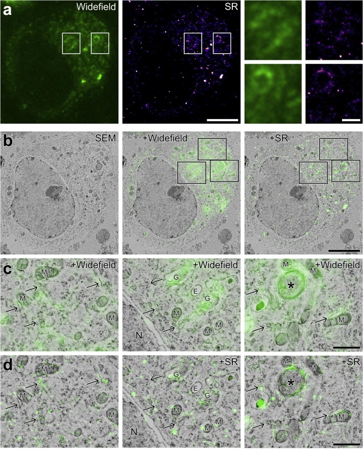
One of the most pivotal innovations is super-resolution light microscopy, which is able to surpass the diffraction limit of light to create images of organic samples at an extremely high resolution. A newly published paper in the Journal of Stuctural Biology attests to the value of super-resolution CLEM with highly detailed images of fluorescent proteins and cellular structures using Delmic's SECOM Super-Resolution system.
According to the authors, a major advantage of an integrated CLEM system is the ability to create an image of a sample with two microscopes simultaneously, which reduces unwanted changes to the sample in between measurements. Although CLEM presents its own unique challenges in terms of sample preparation, the authors demonstrate a workable method using specific fluorescent probes that can be imaged under both EM and FM.
The authors also illustrate the benefits of using a super-resolution light microscope, which is able to localize regions of interest in a much more precise manner. Images show that not only is the lipid dygliceride (DAG) located in a certain region of the cell, but it can be identified specifically within Golgi stacks, endoplasmic reticulum, and a putative autophagosome.
The entire paper can be read here.
If you would like to learn more about Delmic's SECOM Super-Resolution system, we invite you to download the brochure: