.svg "DELMIC")
.png)


This research investigates the potential for using rare earth elements as imaging probes in bioimaging.
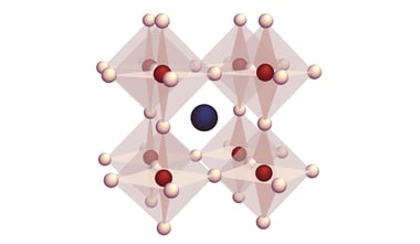
Rare earth elements are known for their luminescent properties. The photoemission of rare earth element-doped inorganic nanocrystals is not severely affected by electron beam exposure, which makes them useful as optical components. The researchers were able to make the luminescent nanocrystals suitable for studying biological samples and thus harmless to cells. Subsequently, they were able to capture high-resolution images of cell cultures.
The research was conducted at the Swiss Federal Laboratories for Materials Science and Technology (EMPA), using the SPARC cathodoluminescence detection system. Traditionally, the SPARC is used for research in the materials sciences, optics, and nanophotonics. However, developments are ongoing as research facilities discover novel uses for our systems.
In related news, last month marked the publication of another paper on the luminescent properties of rare earth materials. This research was also carried out using the SPARC. You can read about it here.