.svg "DELMIC")
.png)


We are excited to announce that, in addition to our upcoming cathodoluminescence webinar, we will also be hosting an informative webinar on the uses and applications for correlative light and electron microscopy (CLEM). At the webinar, we will be providing an introduction to CLEM, featuring our own high-performance CLEM system, the SECOM. Subsequently, various applications for CLEM in the life sciences will be explored and we will highlight a study that demonstrates the potential of correlative microscopy for investigation into type 1 diabetes. We will also talk about one of our latest developments: implementation of large scale imaging using automated data acquisition and stitching.
This webinar is provided by DELMIC’s in-house CLEM specialist, Sangeetha Hari.
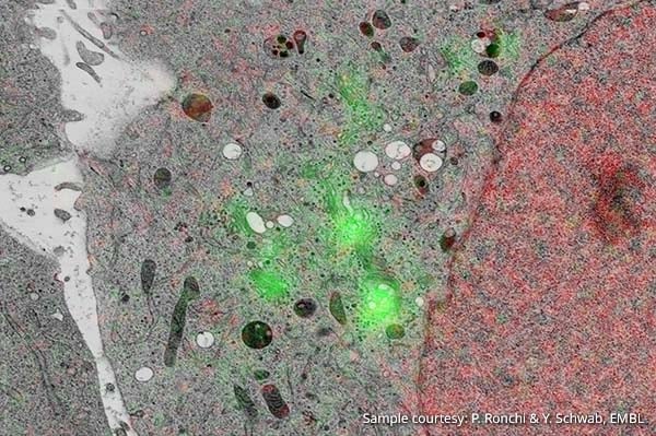
In the life sciences, CLEM is the ideal tool for studying the relationship between form and function of either thin sections of cells or cultured cells. While the electron beam works to create an image that illustrates the detailed structure of a biological sample at an extremely high resolution, the fluorescence microscope that is combined with the SEM to form a CLEM system operates to mark and pinpoint particular elements of the sample. Such an integrated system is ideal for researchers who are concerned with studying samples at a very high resolution while also working accurately and efficiently. We therefore encourage ambitious researchers in the life sciences who want to learn more about how they can improve and streamline their research to sign up for the webinar.
About the speaker
Sangeetha obtained her Bachelor of Science (Honours) in 2005 and Master's degree (2007) from The University of Delhi, India. She went on to work as a Research Scholar in atomic and molecular physics at the Tata Institute of Fundamental Research, Mumbai, India, until 2011, developing time-of-flight spectroscopy systems for the study of electron-molecule collisions and specific bond breaking in molecules. She went on to do a PhD at the TU Delft (until 2016), The Netherlands, to work on a novel technology for sub-30 nm lithography in the Scanning Electron Microscope. She developed imaging techniques to extract quantitative information from high resolution SEM images, using a combination of secondary and backscattered electron imaging with atomic force microscopy. She now works at Delmic as an applications specialist in correlative light and electron microscopy, developing new applications for the SECOM platform.
This webinar is hosted in collaboration with our Australian distributor, AXT.
If you are interested in this webinar, don't forget to sign-up! You can also add the event to your Google Calendar by clicking here. In the meantime, perhaps our application note on type 1 diabetes might interest you: