Their findings have been published in the renowned journal Advanced Energy Materials. The research focuses on CsPbIBr2 which is a fully inorganic perovskite material in which a mix of iodide and bromide is present. It is known that under the influence of external stimuli such as light the I and Br halogen ions can move within the material leading to phase segregation. Such segregation can have a major impact on the electrical properties of the material and its function as solar cell. This effect occurs on microscopic length scales which means that high-resolution microscopy techniques have to be used to investigate it. This particular material is more robust than partially organic perovskite materials making it more suitable for microscopic optical and electron beam studies. As such it acts a good model system for perovskite materials in general. The electron microscopy studies, including the cathodoluminescence (CL) measurements, were performed at the Monash Centre for Electron Microscopy (MCEM).
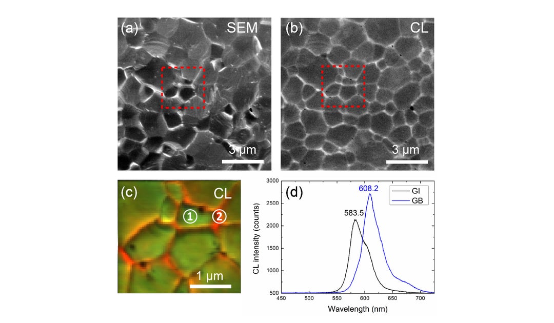
For the CL imaging the beam was raster scanned over the surface and an intensity map was collected. This map reveals that the grain boundaries are brighter.
By performing hyperspectral CL imaging in which a full emission spectrum is measured at every scanning pixel, changes in the emission spectrum can be visualized from point to point. By comparing the grain boundaries with the grain interiors the researchers found that that the boundaries are more rich in Iodide leading to brighter emission and a redshifted emission spectrum. This phase segregation was corroborated by high-resolution TEM studies. Interestingly these iodide rich boundaries dominate the optical response when studying the system with light. Being able to visualize this phenomenon at these small length scales is critical in understanding and quantifying the effect. This knowledge can be used to mitigate adverse effects that are being caused by the segregation and possibly even harness positive effects to improve the cell performance.
The full article can be read here. For questions, you can contact our application specialist Dr. Toon Coenen.
.png)


