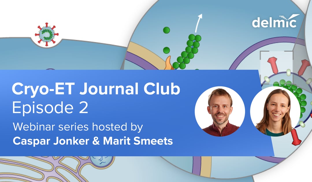
We are introducing the second episode of our new journal club, where we discuss leading cryo-electron tomography (cryo-ET) publications. The emergence of the severe acute respiratory syndrome coronavirus 2 (SARS-CoV-2) has led to a pandemic, causing over 11 million cases and 500 000 deaths worldwide. The world’s scientists and global health professionals are trying their best to look into coronavirus and develop new standards to contain the spread of the coronavirus pandemic and help those affected. In the upcoming discussion of our journal club we will shed light on the articles about how cryo-ET can be applied to study coronavirus.
Our application specialists Marit Smeets and Caspar Jonker will discuss two new preprints on bioRxiv, in which the structure and replication of coronaviruses are characterized using in situ cryo-ET in combination with FIB milling. By using this technique the viruses were studied in great detail during an infection and based on these results both preprints suggested possible drug targets. During the journal club we will evaluate the techniques used and the implications of the findings in the two preprints.
Register now to participate in a live discussion!

30th of July, 10 am (CEST)
We invite you to actively participate in the discussion and share your thoughts in the chat box during the live session. You can read the two preprints and think about the questions below prior to the journal club:
A molecular pore spans the double membrane of the coronavirus replication organelle
SARS-CoV-2 structure and replication characterized by in situ cryo-electron tomography
- The two manuscripts seemingly disagree on the presence of pore complexes in the double membrane vesicles (DMV’s) that house the viral RNA during virus replication. Where Klein et. al. did not find any, Wolff et. al. documented a complete structure. Possible explanations for this could be that the pores are not present at all times during infection, or are not present at all in certain cell types, because there is a separate mechanism. Do you think the pores found by Wolff et. al. are the long sought-after method of RNA export from the double membrane vesicles?
o Yes, the pores are the mechanism for RNA export from DMV’s
o Yes, but there may be additional mechanisms
o No, the pores are not for RNA export from DMV’s
- Do you think cryo-ET will ultimately play an important role in the establishment of a usable SARS-Cov2 drug or vaccine target?
o Yes
o No
If you would like to stay updated about the upcoming episodes, subscribe to the whole series now.

In the first episode we reviewed the article – Ribosome-associated vesicles: A dynamic subcompartment of the endoplasmic reticulum in secretory cells – which was published in Science Advances this year. The authors made excellent use of the diverse capabilities of the cryo-ET technique. You can now watch the recorded first episode below.
This work is supported by the European SME2 grant № 879673 - Cryo-SECOM Workflow