Studying marine microbes, which cover more than 70% of the earth and represent the world's largest ecosystem, can reveal information about ocean's chemistry and climate, and maybe even bring us closer to understanding how the life originated on our planet.
Studying something as tiny as marine microbes can be extremely challenging. Microscopy proves to be a great tool for examining physiological and metabolic states of individual cells. Two types of imaging can be extremely effective. Fluorescence microscopy helps to image the function and provides information regarding metabolism of the organism. On the other hand, it is impossible to get information about ultrastructural composition of the sample. In order to identify cells and different communities of microbes a high resolution imaging, performed by electron microscopy, is required.
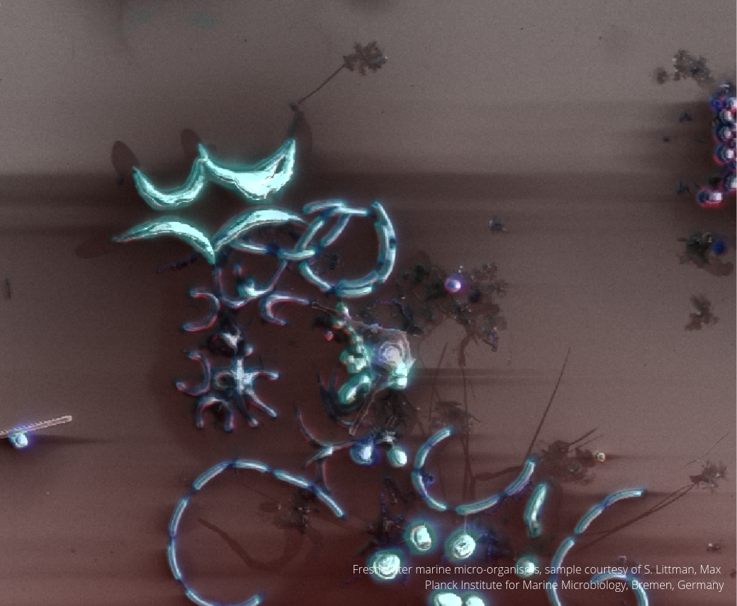
The SECOM combines both of the imaging techniques in one integrated system. With the use of the SECOM it is possible to perform simultaneous correlative light and electron microscopy (sCLEM). Both of the images can be acquired simultaneously without transferring the sample. For the researchers at Max Planck Institute for Marine Microbiology this technique is extremely useful as they are studying marine microorganisms found in the surface water of the Baltic Sea.
To learn how the workflow was performed and how the sample was prepared, read our application note.