.svg "DELMIC")
.png)


We are excited to present a solution that will be able to help researchers and facilities image hundreds or thousands of sample sections 100 times faster and to really understand intermolecular connectivity at the mesoscale.
The ability to image large areas of biological tissue within a short time span will open up many new avenues of discovery, not only in fundamental research but also in drug targeting and potentially even personalized medicine.
Elizabeth Carroll, Assistant Professor at TU Delft: “EM remains the gold standard method to resolve the structure of the synapse. In connectomics, we need to image tens of thousands of thin sections to reconstruct all the connections in a neural network. FAST-EM will make it possible to image more brains so we can start to compare individual animals”.
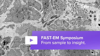
Watch the demonstration of FAST-EM
During the online launch our applications specialist went through the specifications of the system, showed images acquired with FAST-EM and the hardware. The recording is now available for you to watch below!
If you have any questions about the system after the demonstration, please feel free to contact our applications specialist Job Fermie.