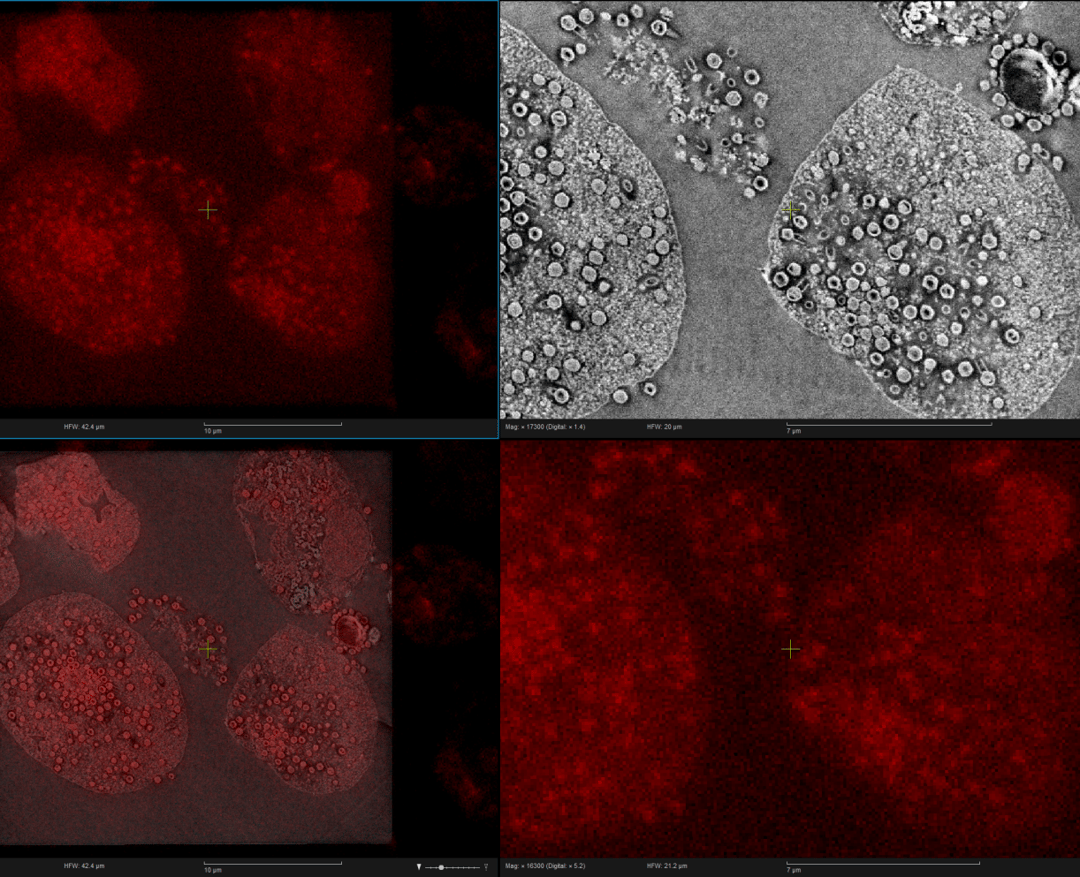
The application note is based on a newly published paper Correlative light-electron microscopy of giant viruses with the SECOM system.
In this research, one type of GVs, Tupanvirus, was studied with correlative light and electron microscopy (CLEM), which is able to show the ultrastructure of fluorescent labeled cells and provide a link between the cellular dynamics and the ultrastructure of any object of interest. The SECOM system allows to perform the light and electron microscopy images without the need of sample transfer and image alignment. With the SECOM, it was possible to successfully detect giant virus particles by fluorescence light microscopy (LM) and simultaneously acquire information with the electron microscopy (EM) ultrastructure such as the Tupanvirus capsid and tail features. This demonstrates that the SECOM can used for screening suspicious samples and virus-infected cells at low and higher resolution, which makes it an extremely efficient tool in the virology field. It describes the benefits of correlative light and electron microscopy for understanding Giant Viruses (GVs), astonishing microbes that are larger than regular viruses. GVs particles and/or sequences can be found in environmental, animal, as well as human samples.
If you would like to read in more details how the sample was prepared, and which other materials and methods were used, make sure to download the application note.
.png)


