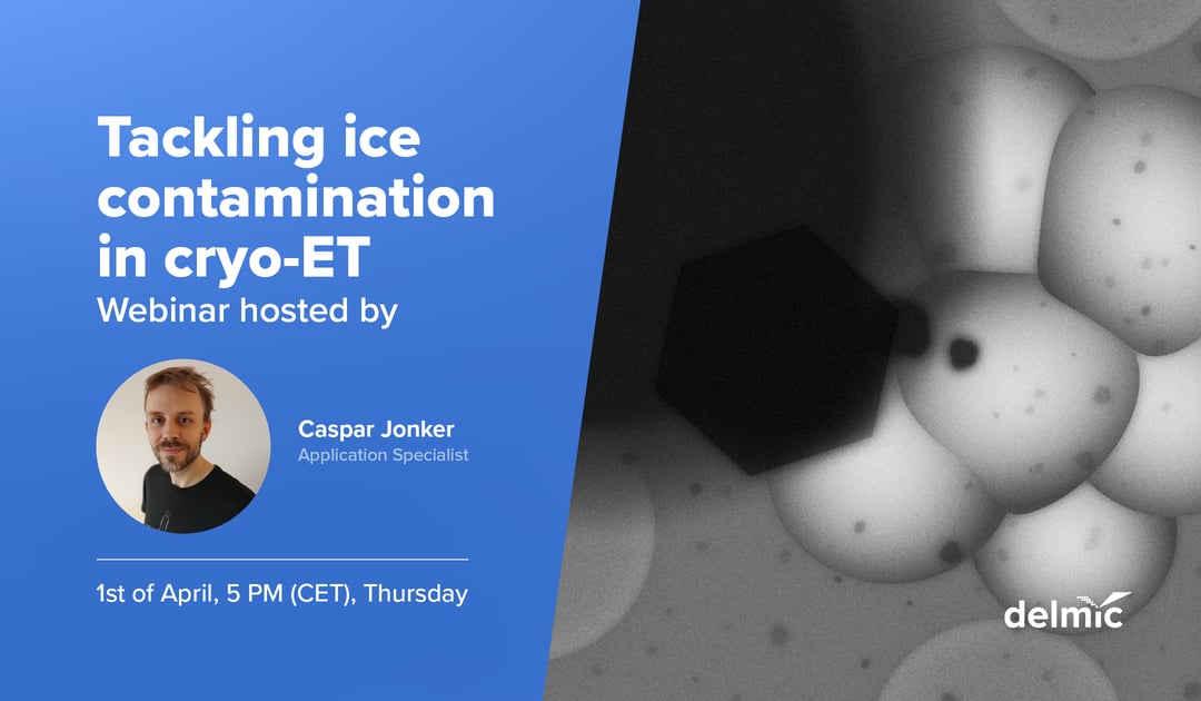
One is the contamination with ice crystals during transport of the sample and the other is contamination occurring even in the high vacuum of the focused ion beam (FIB) / scanning electron microscope (SEM) chamber. Watch the recording of the webinar we held in April to find out the best solution to minimize ice contamination in cryo-ET.
Samples must remain in a vitrified state at ~ -160 °C in the cryo-ET workflow. However, ice contamination of the sample has been a common and severe challenge hindering the progress of cryo-ET workflow. One of the two major contamination sources is the contamination with ice crystals during transport of the sample. Samples transported or handled in liquid nitrogen can easily be contaminated with ice crystals that have formed when the liquid nitrogen, or the tools that are used in liquid nitrogen, have been exposed to the open air. Samples transported in low vacuum can also attract water molecules that form crystals on the surface of the sample. Ice crystals obscure regions of interest, hampering the acquisition of meaningful tomograms. The second major contributor to contamination occurs even in the high vacuum of the FIB/SEM chamber. Here, water molecules can deposit and form a crystalline ice layer on the lamellae at a rate of around 50 nm per hour. With the process of lamellae milling lasting many hours, this can cause a thin, ~150 nm lamella to be substantially thicker after all milling is done. The thick crystalline ice layer substantially reduces the quality of the acquired tomogram.
In this webinar our application specialist Caspar Jonker is going to introduce the crucial steps in the workflow that contribute most to sample ice contamination and most importantly dive into the new developments that aim to minimize contamination in cryo-ET altogether. Watch the recording now!
.png)


