.svg "DELMIC")
.png)


Are you joining Microscience Microscopy Congress 2021 this year? Even though it is taking place virtually this year, the participants will be able to join multiple meetings and workshops. The event will take place from 5th until 9th July 2021.
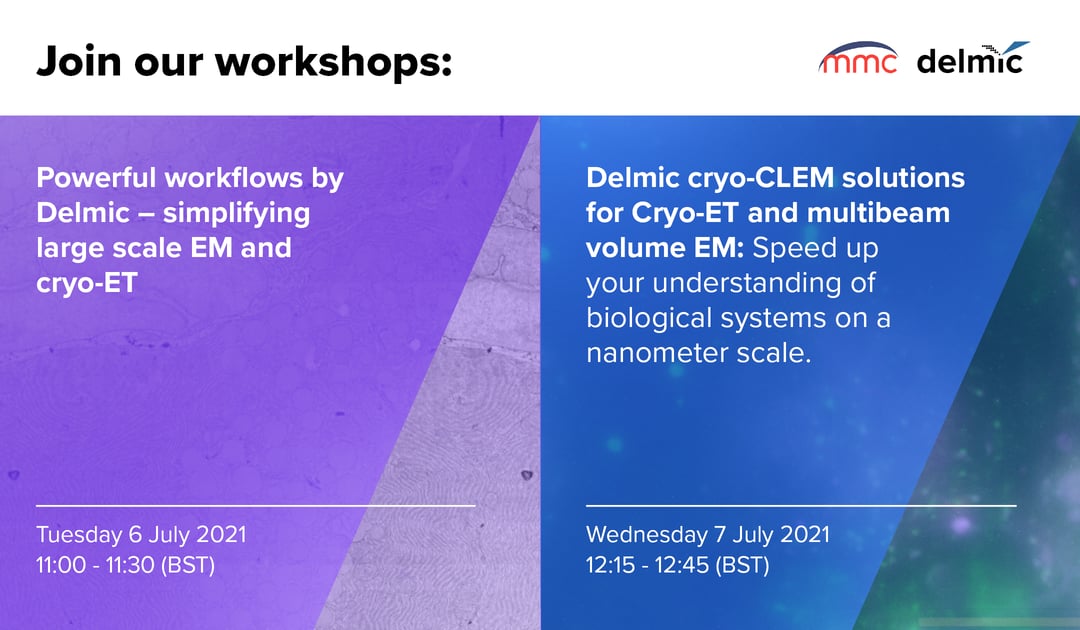
We will be presenting our workflows during two exhibition workshops, which will be free to attend by anyone. Make sure to reserve your spot!
Powerful workflows by Delmic – simplifying large scale EM and cryo-ET
In this workshop, we will discuss how our METEOR and ENZEL systems can boost your success with cryo-electron tomography (cryo-ET) through gaining integrated cryo-CLEM capabilities. Secondly, we will explain how our FAST-EM multibeam volume EM system helps you get detailed molecular information and context from large biological samples 100x faster.
Cryo-ET uniquely provides high-resolution structural information on biomolecules within their native environment. However, the technique requires a challenging lamellae milling process using a focused ion beam (FIB). The lamella fabrication yield is currently dampened by the difficulty in region of interest (ROI) targeting as well as ice contamination and devitrification of the sample during sample transfer. In this workshop, we will show how our integrated cryo CLEM solutions guide lamella milling in situ, increase milling precision and allow for ROI inclusion verification. We will explain how integrated cryo-CLEM streamlines the cryo-ET workflow, and increases sample yield with examples from yeast cells, HeLa cells and Dropsophia myofibrils.
Electron microscopy can often represent a bottleneck in the quest for answers to various scientific questions in fields such as cell biology, pathology, toxicology and neurobiology. The limited throughput of current technology prevents imaging of large volumes of biological material in reasonable timeframes, hence relegating EM to providing only qualitative information. FAST-EM enables a shift towards using EM as a fast quantitative analysis tool. Higher throughput drastically shortens the time needed to acquire project data, and opens the door for projects of unprecedented scale, which were previously too time-consuming to consider. We will demonstrate high-throughput imaging of a variety of tissues and experimental systems, showing how high-resolution data can be placed within a large-scale context.
Time and date: 6th of July at 11:00 (BST) (free to attend)
To learn more about registration, please check this page.
Workshop: Delmic cryo-CLEM solutions for Cryo-ET and multibeam volume EM: Speed up your understanding of biological systems on a nanometer scale
In this workshop, we will present our most recent solutions to simplify cryo-electron tomography (cryo-ET) and large-scale EM.
Cryo-ET is an imaging technique that allows reconstruction of high-resolution models of protein complexes in their near-native state. However, the current workflow is error-prone resulting in damaged, contaminated and devitrified samples that cannot be used for data analysis. To overcome these problems, we developed METEOR an integrated cryo-CLEM system to guide cryo-lamella milling and ensure the region of interest is present in the final lamella. We will also present CERES, our ice defence system minimizing ice contamination in every step of the workflow. This will allow researchers to obtain better quality cryo-EM and cryo-ET results, higher throughput, and higher resolution.
Large scale EM aims to integrate nanoscale observations into the larger context of the sample, which can cover a large area or span through a three-dimensional volume. With conventional large-scale EM workflows, operators are forced to balance the size of the imaged area against the resolution needed to visualize the required level of detail. Despite best efforts, large projects still take days to months of continuous imaging when high-resolution data is needed. To overcome this challenge, we have developed FAST-EM, an automated multibeam scanning EM, which combines simultaneous 64-beam electron imaging with an automated workflow. This enables users to tackle current and future large-scale projects such as volume-EM of cells and tissues, connectomics, and large-scale 2D nanotomy projects within hours or days instead of weeks or months.
Time and date: 7th of July at 12:15 (BST) (free to attend)
To learn more about registration, please check this page.
To learn more about Microscience Microscopy Congress please visit the official website.