.svg "DELMIC")
.png)


A paper on the benefits of the integration of electron and fluorescence microscopy has recently been accepted to be published in ACS Nano. High-resolution imaging of live cells at the scale of a few nanometers is always a challenge because of the damage caused to cells by the process of electron microscopy, by which cells are fixed or frozen and placed in a vacuum. Research conducted at the Department of Imaging Physics at Delft University of Technology and the Department of Molecular Physiology and Biophysics at Vanderbilt University explores this challenge through an innovative method for imaging live cells with both fluorescence and electron microscopy. DELMIC’s Daan van Oosten Slingeland was one of the contributors of the project.
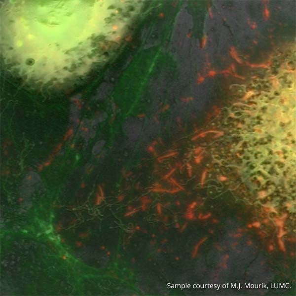
This paper presents the ability for a correlative system to identify the relevant sites with FM prior to exposure with an electron beam. The dynamic nature of fluorescence imaging allows one to identify certain elements at particular moments as they are occurring in time before they are potentially altered or destroyed by the effects of electron imaging. Once the site has been identified, it can then be imaged with an SEM which provides structural information and increases the resolution 30-fold.
This was demonstrated by analyzing the uptake and transport of bio-conjugated quantum dots on cellular extensions. Quantum dots are highly efficient tools for analyzing samples with fluorescent light due to their unique optical properties. The quantum dots were identified as they were actively being transported through the cell with fluorescence imaging. The electron microscope then provided more high-resolution details of the context and structure in which they are situated.
This article attests to the importance of using a correlative system for obtaining detailed information about organic samples with consideration for the region and moment in time when the images are obtained. An online version of the article can be found here.