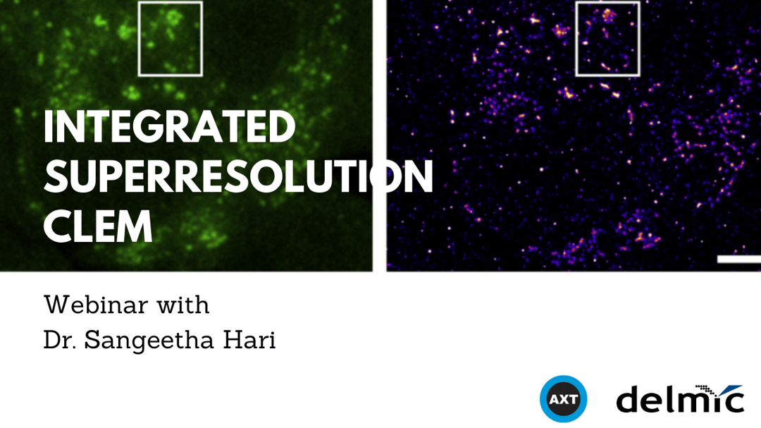
In the webinar, our application specialist Sangeetha Hari presented our SECOM SR system which is an integrated platform for CLEM that uses super resolution optical microscopy in combination with a scanning electron microscope to perform SR-CLEM. The principle of operation, details of CLEM, hardware, open source software-based operation and scripting, as well as sample preparation were discussed in this webinar. For those who have missed the opportunity to watch the webinar live, it is now available on our website for free.
Correlative light and electron microscopy (CLEM) is a powerful imaging technique for applications in life sciences. It enables the study of function and structure at the sub-cellular level due to the combined ability of fluorescence and electron imaging. Integrated correlative microscopy (iCLEM) combines these two methods to offer functional information in the context of high resolution structural information, while allowing the imaging to take place in one system without the need for sample transfer between microscopes or external fiducial markers.
In this webinar we present a super resolution (SR) imaging system integrated with a scanning electron microscope to perform SR-CLEM, achieving a resolution down to 85 nm, imaging in-resin fluorescence from GFP in thin sections. The principle of operation, details of CLEM, hardware, open source software-based operation and scripting, as well as sample preparation will be discussed in this webinar.
This was our fifth webinar this year and we anticipate to be hosting more webinars in the near future. We would like to extend our thanks to Dr. Sangeetha Hari - Delmic’s correlative microscopy application specialist – for giving the talk, and our Australian distributor AXT for their efforts in hosting the webinar.
.png)


