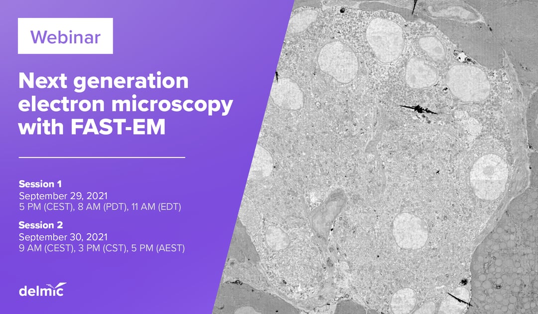
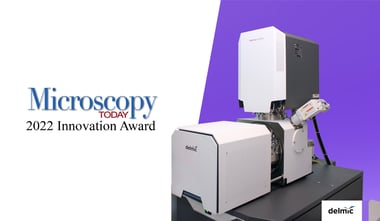
If you answered 'Yes' to any of the above questions, then we invite you to join our upcoming webinar: Next generation electron microscopy with FAST-EM. The webinar will take place on the 29th of September at 5 PM (CEST) and on the 30th of September at 9 AM (CEST). Together with our speaker and applications specialist at Delmic Job Fermie we will show a full walkthrough of the FAST-EM, our new multibeam scanning electron microscope.
The system was launched at the end of last year, and now we are excited to show its full capabilities. FAST-EM can image biological thin samples at unprecedented speeds, and with a level of automation that enables large scale imaging without the need of constant supervision.
During this webinar we will go through all the steps that are required for capturing large-scale data, including:
- sample preparation
- sample exchange and loading
- creating overviews and using them to acquire data
- image acquisition
- browsing through acquired data
We will also share a link where you will be able to navigate the resulting high-resolution images yourself. In the end, we will discuss future possibilities and applications, and answer your questions.
Reserve your spot in one of the two available sessions to be the first one to see FAST-EM in action.
.png)


