.svg "DELMIC")
.png)


The paper, published in Journal of Microscopy, explains the new method of automated data acquisition for integrated light and electron microscopy. More specifically, the authors present a scheme for automated detection of fluorescent cells within thin resin sections for following electron microscopy imaging. This method aims to speed up the process of data acquisition and reduce the data rates.
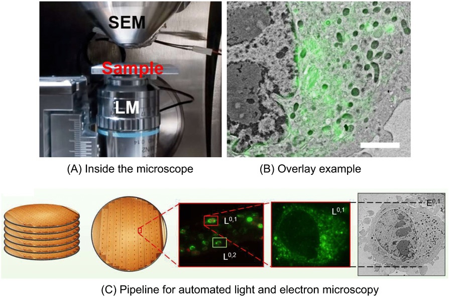
Correlative light and electron microscopy or CLEM combines the power of light and electron microscopy and provides functional information in high‐resolution. Array tomography, a method of imaging ultrathin sections cut through resin‐embedded cells, can be performed inside of a light and electron microscope to acquire a high-resolution and functional data. One of the difficulties of this technique is the enormous amount of data produced, which takes a lot of time and effort to analyze. The authors propose a new method of automated array tomography inside of the light and electron microscope, which has a potential of reducing the volume of data by marking the regions of interest.
The imaging was performed on SECOM light microscope platform, a unique solution for extremely fast correlative microscopy with the highest optical quality and overlay accuracy.
You can read the full article here. If you would like to know more about correlative light and electron microscopy, check our resource library.