.svg "DELMIC")
.png)


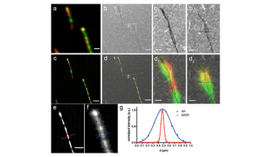
Superresolution (SR) correlative light imaging technique provides highly accurate correlation of fluorescent proteins with cellular structure. The new application note focuses on the benefits and possibilities of The SR-iCLEM (superresolution integrated CLEM) technique.
The major advantage of this technique is the simultaneous correlative image acquisition, which allows to avoid sample contamination and difficulties in the image overlay. In the application note the overlays of the WF-EM and SR-EM images are compared. While the fluorescence could be assigned to individual virus particles, based on the WF images, it is only from the SR reconstruction that it can be seen to originate from the core of the virus. Therefore, the combination of the SR and EM imaging allows to assign the SR signal to specific organelles.
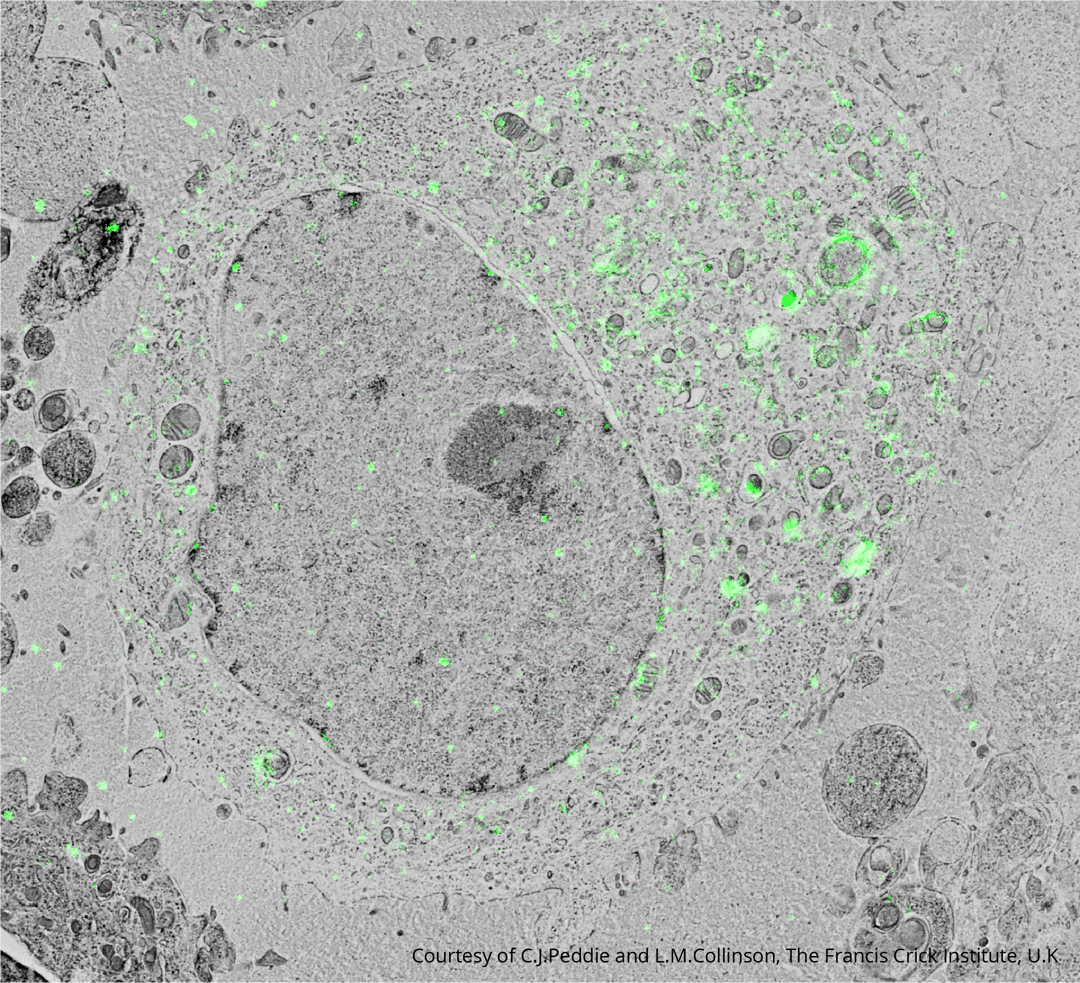
The imaging was performed with the SR SECOM system in Francis Crick Institute, you can read more in the SR SECOM brochure. We invite you to download the application note here.