.svg "DELMIC")
.png)


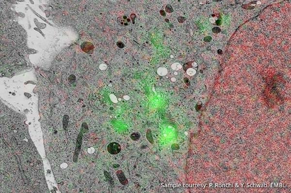
These images are overlayed to obtain information on proteins or small organelles within their ultrastructural context. The most common method to achieve overlay is with the use of fidicual markers. When dealing with a nanoscale resolution, however, fiducial markers can be cumbersome and unreliable. A paper published in the Scientific Reports section of Nature.com demonstrates an alternative method which is reliable and straightforward: automated overlay.
A unique patented cathodoluminescence marker technology, developed by Delmic, makes overlay a seamless and automatic procedure. This is only possible in an integrated system which combines a SEM with a fluorescence microscope (iCLEM): the cathodoluminescence pointers from the electron beam are directly visualized in the fluorescence detection channel. This results in highly accurate data free from user interpretation. In this paper, the authors show that this method allows for the acquisition of combined data at a resolution of 5 nanometers.
We at Delmic are pleased to see a growing recognition for the benefits of automatic overlay. This solution is commercially available as the SECOM: a correlative light and electron microscopy system which fits on a scanning electron microscope.
In the biological sciences, researchers often rely on the combined data from an electron and fluorescence microscope.