.svg "DELMIC")
.png)


Inspired by the temporary exhibition Unimaginable in the Rijksmuseum Boerhaave
It was on a Thursday afternoon in June when I came across an extraordinary story at the Rijksmuseum Boerhaave in Leiden [1]. I had heard about a temporary exhibition on microscopy in this science and medicine museum and hoped it would unveil interesting stories worth sharing. So, my colleague and I took the train from Delft to Leiden and spent over three hours exploring everything there was to see.
 The temporary exhibition 'unimaginable' at the Rijksmuseum Boerhaave. Image taken from [1].
The temporary exhibition, titled ‘Exhibition Unimaginable’, centered around a man named Antoni van Leeuwenhoek, who was born nearly 400 years ago in Delft, a city in the Dutch Republic. He grew up in the middle of the ‘Golden Age’, a period marked by a renaissance of advancements in science and art. Little did he know that his groundbreaking discovery of bacteria would still be celebrated after four centuries. This is his remarkable story.
Van Leeuwenhoek was not a scholar with a university degree, nor did he belong to the ranks of nobility or wealthy citizens who pursued science as a hobby. Instead, he was the owner of an ordinary draper shop in Delft. However, he developed an interest in lens making, possibly as a means to better observe the quality of cloth [2]. He devised his own method for creating very powerful small lenses and made single-lens microscopes capable of magnifying objects up to 250 times or even more [2].
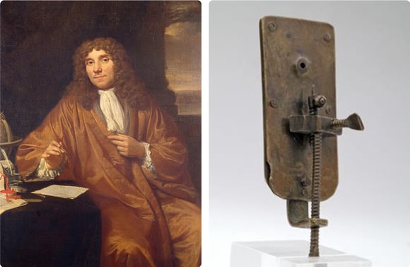 Left: Antoni van Leeuwenhoek, right: Single-lens microscope made by Antoni van Leeuwenhoek. Taken from [1].
Driven by curiosity
At this time, around the year 1670, microscopes had already been invented and were gradually being used for scientific purposes. However, achieving a magnification of 250x was unprecedented for its time. Van Leeuwenhoek could have simply used his powerful single-lens microscope to examine cloth, and that would have been the end of this story. Instead, he got very curious.
He likely found inspiration in the book ‘Micrographia’ by Robert Hooke, published in 1665 [3][4]. Hooke, a member of The Royal Society in London, was among the first to use a (compound) microscope for studying objects and living organisms. The book displayed very detailed drawings of small objects such as hair, organisms like fleas and ants, and even microfungi - the first microorganisms ever observed.
Van Leeuwenhoek started to use his one-lens microscope to observe the world. His curiosity drove him to look through the microscope at various objects, such as his own beard hair, sperm cells, molds, and lice. Subsequently, he would write letters to the Royal Society of London, explaining his findings. In total, he wrote approximately 200 letters [3].
Discovering the microworld
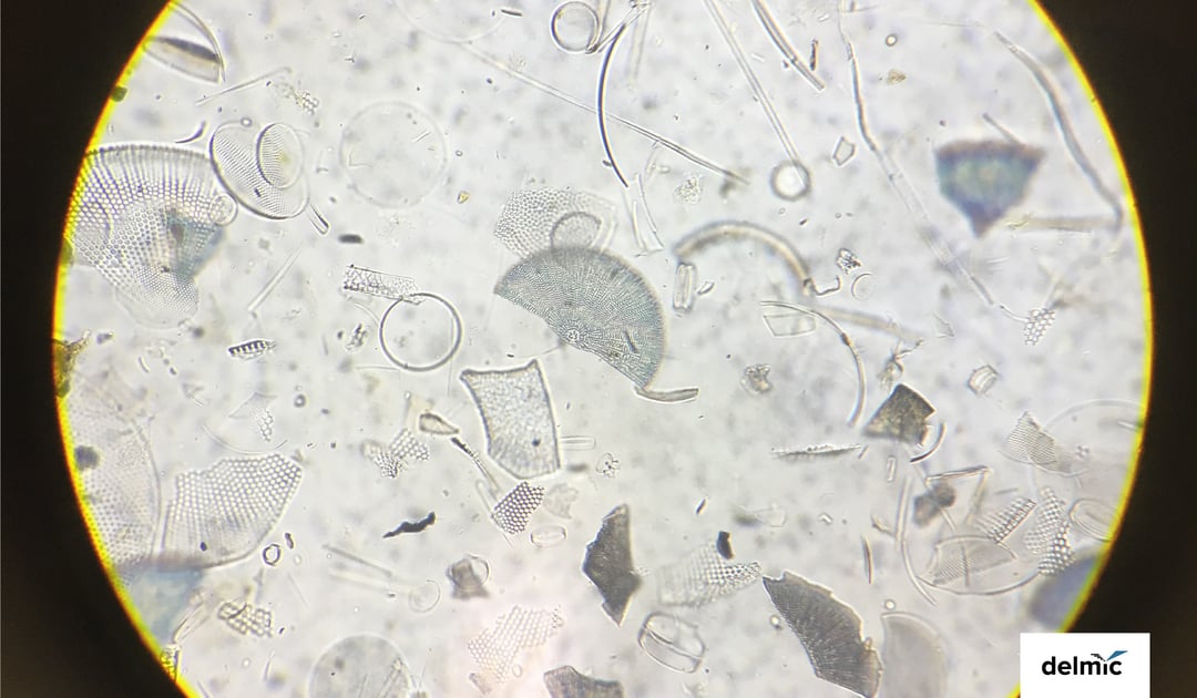
In 1674, Van Leeuwenhoek wrote a letter describing an experiment where he investigated water from the Berkelse Mere, a lake in Delft. At the time, nobody thought it would be interesting to look at water under a microscope. However, when Van Leeuwenhoek did, he was amazed at what he was seeing. With his tiny microscope, he saw that thousands of small creatures were swimming around in the drop of water.
He describes little green clouds he observes, and many little ‘animalcules’: “The motion of most of these animalcules in the water was so swift, and so various upwards, downwards and round about that ‘twas wonderful to see: and I judged that some of these little creatures were above a thousand times smaller than the smallest ones I have ever yet seen upon the rind of cheese” [3].
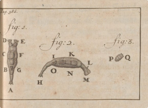 Figures of tiny 'animalcules' that Antoni van Leeuwenhoek observed. Image taken from [1].
So what did Van Leeuwenhoek actually observe in the water? He observed protists and bacteria, probably ciliates and rotifers [5]. However, the specific types he saw are still debated among microbiologists today [3]. This uncertainty stems mostly from his cryptic descriptions and translation errors in his letters [6]. Nevertheless, it's undisputed that Antoni van Leeuwenhoek was the first to observe single-celled organisms, including bacteria.
Convincing the scientific community
Since Van Leeuwenhoek was the only person who could observe these small ‘animalcules’, the Royal Society was suspicious, and people accused him of telling fairytales [3]. Furthermore, his lack of formal education made it challenging for him to convince the Royal Society of the validation of his findings.
Over the following years, Van Leeuwenhoek continued to observe and document his findings. His letters were often read out loud and started to attract great interest. He gathered testimonials of his findings, although some scholars, like Christiaan Huygens, remained skeptical. Fortunately, Robert Hooke could eventually replicate his experiments, providing validation and leading to Van Leeuwenhoek’s election as a fellow of The Royal Society in 1680 [2]. He received the credit he deserved.
Back to the present day
As I listened to this story in the museum, I couldn’t help but wonder what it must have been like to be the first person to see bacteria in a drop of water. Van Leeuwenhoek himself vividly describes his amazement when discovering these tiny organisms. Fueled by his curiosity, he explored the microworld with an open mind, and his efforts eventually paid off.
Personally, my curiosity also drove me towards science, and I’m still amazed when I read about new discoveries and innovative techniques. However, as I walk through the museum, I’m captivated by the interesting history of science and medicine. From the theatres where people used to study cadavers to the first laboratories, from the first microscope to techniques like cryo-electron microscopy (cryo-EM), we have definitely come a long way.
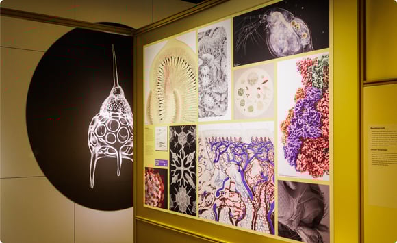
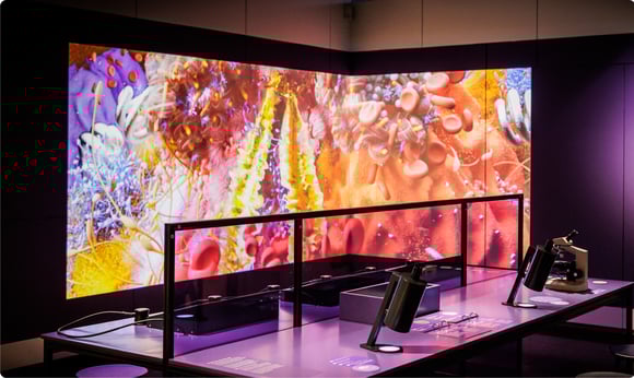 The temporary exhibition 'Unimaginable' displaying the current state of microscopy. Image taken from [1].
To show this progression, the temporary exhibition also displays a video about the study of the Treponema denticola bacterium. Antoni van Leeuwenhoek was the first one to see this spiral-shaped bacteria, and recently, the mechanism of its movement was revealed using cryo-EM. Vera Williams, a scientific visualizer, made a beautiful animation of this bacteria and its mechanisms [7].
After the museum closes, we take the train back to Delft again. I feel inspired by the history I’ve come across, and I think about the new technologies and scientific discoveries we will experience in the future. Where will the future lead us?
References
[1] Rijksmuseum Boerhaave, Leiden
[2] Gest, H., Notes and Records of the Royal Society of London 58, 2, 187-201 (2004)
[3] Lane, N., Philosophical Transactions of the Royal Society B: Biological Sciences 370, 1666, 20140344 (2015)
[4] Micrographia by Robert Hooke, Google Books
[5] Dobell, C., Antony van Leeuwenhoek and his ‘little animals’, Harcourt, Brace and Company (1932)
[6] Van Egmond, W., Micscape Magazine 239 (2016)
[7] Vera Williams, scientific animations