.svg "DELMIC")
.png)


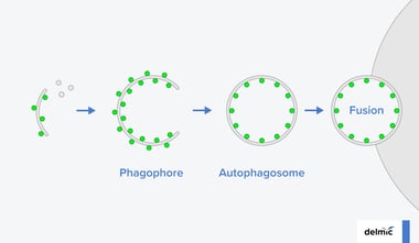
Light or fluorescence microscopy made it possible for the researchers to detect the functional information and image different colors and parts of the cell. It provides the data to understand the dynamics of the cell, however, the diffraction limit of light doesn’t allow distinguishing objects that are smaller than the wavelength of light. That is when the life scientists turn to electron microscopy, which provides the structural information in a high resolution.
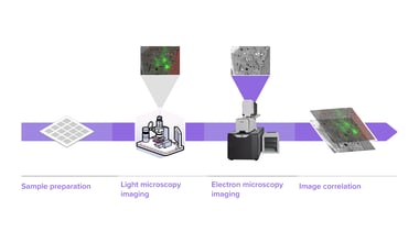
It is very common to use more than one method to get the data about the sample. Using two different modes of microscopes usually means a lot of challenges. First of all, it takes a lot of time to process the sample from fluorescence microscope and transfer it to the electron microscope. Secondly, overlaying the images is extremely difficult and often results in poor data accuracy. Finally, in the process of transferring the sample from light microscope to electron microscope, there is a chance of contamination or even damaging the sample completely.
Correlative light and electron microscopy (CLEM) combines the advantages of both techniques and helps to obtain the results faster and easier. Together with our application specialist Sangeetha Hari we prepared a video, which explains correlative microscopy in the details.
SECOM is the integrated correlative light and electron microscopy solution, which allows performing correlative microscopy extremely fast with the highest optical quality and overlay accuracy. To read more about the research done with the SECOM system in the spheres of biology, neuroscience and cancer research read our application notes.