This article illustrates the usefulness of a correlative approach for biology, as it provides the ability to analyze the same samples at varying length scales, to alternate between different microscopes without compromising the sample, and to ensure optimal overlay of images.
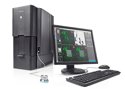
In biology, the Delphi system is ideal for studying resin -embedded tissues or cells, mono-layers of whole cells, and isolated particles on a substrate. For this paper, the Delphi system was tested by analyzing monolayers of HeLa cells labelled with fluorescent markers for endosomes and for the actin cytoskeleton. The system was successful in imaging the same cells with its electron and fluorescent microscope. These images were then automatically overlaid by the Delphi software.
Sample preparation is of essence and can be a challenge, as many factors have to be taken into account when analyzing the various aspects of a sample in correlative imaging. However, the Delphi system has demonstrated that there are methods in which samples can be prepared to be optimal for both FM and EM imaging. The Alexa fluorophore dye used in this particular test case remained active after drying (which is required for imaging in a vacuum), at the same time that ultrastructural details could be identified with the electron microscope. It is anticipated that more sample preparation protocols will be developed in the future in order to ensure that researchers from a wide variety of disciplines can benefit from the efficient correlative system offered by the Delphi.
To read this edition of Microscopy & Analysis, click here. The paper can be found on pages 25- 29. For any further inquiries, contact Lenard Voortman at voortman@delmic.com.
.png)


