.svg "DELMIC")
.png)


While electron microscopes and fluorescence microscopes have been go-to techniques for studying organic samples at a high resolution, individually they fall short in offering the exhaustive data needed for truly in-depth life science research.
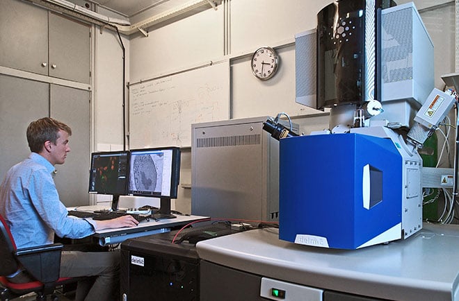
In 2011, the Charged Particle Optics group at TU Delft completed the development of the SECOM. This system integrates a light and electron microscope, thus combining the labelling capabilities of fluorescence microscopy with the high-resolution nanoscale data obtained from electron microscopy. Six years later, the department of Imaging Physics houses no less than five SECOM systems.
We went to visit Jacob Hoogenboom, a central player in the invention of the SECOM and a researcher at ImPhys, to investigate the many applications for which these systems are being used today. The SECOM, itself a pivotal innovation in microscopy, is being used for further studies into new ground-breaking imaging technologies.
Due to its modular design, scientists at ImPhys were able to create a total of five different microscopy systems using the SECOM: a standard CLEM system, a confocal system, multibeam SEM, time-resolved cathodoluminescence CLEM and a system for testing new components and analyzing fluorescence in vacuum.
Confocal light microscopy for thicker samples
The very first SECOM system is now housed on an FEI Verios scanning electron microscope. Originally a widefield microscope, it is now being customized to be a confocal microscope, thus reducing the out-of-focus light that is seen through a widefield microscope. To do this, Jacob’s group is using a standard confocal unit that is fitted on the SECOM door with a separate adaptor module. The inside of the SECOM remains unchanged. This is done with the end-goal of determining an effective method for measuring thicker samples.
Jacob explains, “Ultimately, we want to be able to visualize live cells in a microfluidic environment; we’ve already published some Proof of Principle experiments with the widefield SECOM last year in ACS Nano. The idea is that you follow the cell and the dynamics within the cell using the fluorescence microscope, and then determine the timeframe and a region of interest for when you take the EM snapshot. This is not normally possible with living cells, but because you are able to localize the timeframe and region of interest via the fluorescence microscope, you can get the information that you need without being hindered by the damaging effects of the electron beam on the cell. This is only possible when a fluorescence microscope is integrated with an electron microscope.
“The problem with the widefield microscope is that you need to image through the fluid to get to the region that you want to capture with the electron microscope. We are working with a distance of approximately 100 micrometers, and the liquid is about 70 micrometers thick. So in these first experiments, the correlation and precision was sub-par, and we had issues with background signal. This is something we are hoping to overcome with a confocal system.
“For the first Proof of Principle that we’re working on, we want to use a microfluidic holder with fixed cells. This way, we don’t have to measure in liquid or using live cells. This allows us to turn the cell over so that we can observe the adhesion side of the cell instead of just the top.”
Testing fluorescent dyes with a focused ion beam
Jacob then led us to a SECOM retrofitted on FEI FIB 200, a focused ion beam system. While the FIB is effectively broken, it is being used for its still-functioning vacuum chamber. This system has been customized in such a way that it operates as a widefield excitation and emission microscope; complete with a breadboard in the back, a camera, and a light engine.
“We are using this to test a series of fluorescent immunodyes, to understand the difference between measuring in ambient conditions and in vacuum. We have found that some dyes work more efficiently in vacuum while some bleach very quickly, for instance.”
Cathodoluminescence-based time resolved experiments
The SECOM also has great potential for scientific fields outside the life sciences. Jacob showed us another experimental setup where cathodoluminescence-based time resolved experiments are being conducted for applications in nanophotonics. This setup features a commercial beam blanker in the SEM which allows Jacob’s group to switch from pulsed to continuous operation of the SEM. Electron pulses of 80 - 90 ps duration are generated by conjugate blanking of the electron beam which allows probing emitters within a large range of decay ranges.
The group ultimately published a paper about a method for achieving both temporal resolution (far below typical emitter lifetimes) and spatial resolution (far below the diffraction limit) simultaneously, while a beam blanking approach is used that only requires standard SEM hardware and electronics.
Moreover, moving beyond cathodoluminescence, the setup features ultrafast laser excitation of the sample via the SECOM. Using dispersion compensation optics, the sample can be excited with 20 femtosecond laser pulses focused in a 620 nanometer sized spot. This gives the possibility to measure non-linear optical responses and relate these to high-resolution structural details measured with the SEM, or to do ultrafast pump-probe experiments.
Multibeam SEM: the next great innovation for biology?
We were then guided to the multibeam scanning electron microscope on which the SECOM is retrofitted as a detector. The microscope can currently emit 169 electron beams at once, with the ultimate goal of emitting 200 beams. A challenge however is to detect the signals from all beams separately. For this, the SECOM is used as a transmission detector. Research is also being done into this transmission versus regular backscatter detection, and findings indicate that both detectors work equally well at low energy. This opens up a world of possibilities for biologists.
Such a system would find applications in large scale imaging and the imaging of serial sections. With such a multibeam-SEM, one could measure volumes as large as several cubic millimeters of a brain, or map an entire tumor cell in a fraction of the time this currently takes.
“This is part of a more general move towards higher resolution combined with higher throughput for new data. Before this, our focus was on developing accurate correlative methods and optimizing the workflow. Now, we are aiming to intensively improve techniques and even develop entirely new technologies.”
If you are interested in learning more about the SECOM, we invite you to download the brochure below: