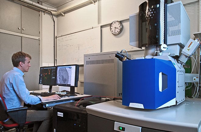
In a labyrinth of options, we offer the technology and the ongoing personal service needed for today's ambitious researchers in the life sciences: the SECOM system, and a team of committed engineers to help you throughout the process of your research.
This article outlines the three main reasons why the SECOM system is the best choice for your research in the life sciences.
1. Automated Overlay
As a company committed solely to developing correlative microscopy solutions, we produce high-performance systems that are fine-tuned to the specific needs of scientists. The most important feature of the SECOM is the straightforward automated overlay provided by the integration of an electron and fluorescence microscope into a single system.
Different overlay techniques are already offered by various manufacturers, but a method has yet to outperform the accuracy and ease-of-use of SECOM's automated overlay. See below a breakdown of overlay methods and how they compare to automated overlay (you can read more about it here):
| Technique | Method | Limitations |
| Universal sample holders | Using a sample holder with markers to indicate the region of interest on the sample | Not exact and therefore not very reliable: highest precision at 10 micrometers |
| Manual overlay | Using image editing software (ex: Photoshop) to combine the images into one | Based on intuition: risk of bias |
| Fiducial markers | Placing physical markers such as fluorescent microspheres in the sample | Requires deep expertise, time-consuming |
With the automated overlay procedure of the SECOM platform, EM and FM images are always aligned perfectly. The unbiased automated overlay feature makes use of the unique patented cathodoluminescence marker technology which ensures highly credible and efficient life science research. The key to this procedure is the physical principle of cathodoluminescence: light is generated at the position where the electron beam hits the glass. This light can be detected by the camera of the fluorescence microscope and acts as a temporary fiducial marker.
The overlay is carried out by ODEMIS: a sophisticated, yet straightforward, software package that is integrated with the system. ODEMIS was designed at the DELMIC headquarters in order to make its systems easy to use and to meet the specific requirements of modern research facilities.
For an integrated system, the SECOM also has exceptional optical performance because it is made from the highest quality components. This leads us to our second point:
2. Unrivalled performance
The SECOM has a modular design that enables it to be fitted easily on any scanning electron microscope. For this reason, the system maintains the integrity of the scanning electron microscope, thus not compromising on EM performance. Detail and accuracy is further ensured by a closed loop sample stage, enabling precise acquisition procedures as well as the possibility to retrace your steps during the measurement process.
The SECOM system is also made of only the highest quality optical components to enable accurate, high-quality images. The SECOM thus ensures the best performance of any integrated system without compromising the quality of the fluorescence or electron microscope. The following features ensure that the SECOM matches the quality of any existing optical microscope:
- Scientific CMOS cameras with a large chip are used, providing an extra large field of view
- Data acquisition across various frequencies with multiband imaging
- Minimal optical aberrations due to full apochromatic and planar correction
- Use of the highest quality filters: your sample is therefore not unduly bleached
The SECOM also makes use of a unique vacuum compatible immersion oil. This allows you to use high-numerical aperture objectives in the SECOM platform. Immersion is a technique which increases the resolving power of a microscope. BitesizeBio explains this very straightforwardly:
With non-immersion (or ‘dry’ as they are called) objectives, there is an air gap between the front lens of the objective and the top surface of the coverslip. Most microscope slides and coverslips will have refractive indexes of 1.5, whereas air has a refractive index of 1.0. And as you know, when light passes from one medium to another, say from glass to air, it ‘refracts’ or bends and scatters. Therefore, if you use a dry objective the light rays from your specimen will undergo refraction when travelling from the glass coverslip into the air. Refracted rays are not usually collected by the objective front lens and are lost to the final image.
Now if you use an immersion medium to replace the air gap you can correct this mismatch. Most of the commercial immersion oils have refractive indexes of around 1.51 – similar to glass. This improves the resolution of your high-power immersion objective and increases your NA by lowering refraction.
As a testament to the high-quality optics that we use to create our systems, we also produce a version of the SECOM system with a super-resolution fluorescence microscope, the SECOM SR, thus offering the best of both worlds in electron and optical microscopy. Super-resolution is able to overcome the diffraction barrier of light, which was previously impossible with optical microscopy.
Super-resolution microscopy allows one to observe small organelles, proteins, and textures. With our super-resolution CLEM system, the electron microscope offers yet more detailed and contextual information to further strengthen the integrity of your research.
Image: Localization of the lipid diacylglycerol within cellular membranes of HeLa cell expressing GFP-C1, imaged using the SECOM system.
Incidentally, the SECOM SR system came into existence as a response to the needs of one our existing customers, which leads us to our final point:
3. User-focused
As we mentioned above, a microscope is a massive expenditure, in terms of funds as well as time. Obtaining the SECOM, however, makes this a worthwhile investment because of the personal contact that we maintain with our users; both during the process of purchasing the system as well as during its actual use.
DELMIC is a relatively fresh company in the world of microscopes, which is largely made up of giants who produce a wide range of consumer products. We are a young group of scientists and engineers who are very familiar with the demands of research facilities and what it means to carry out the most insightful and innovative research in a competitive environment. The SECOM is thus a finished product designed for precise data acquisition in a research facility, at the same time that it can be customized to the specific needs of the researcher. The SECOM also ensures flexibility in terms of your electron microscope: the system is designed to be fitted on any SEM by major manufacturers.
The SECOM SR system became commercially available after we initially produced it for one of our existing users, Dr. Lucy M. Collinson at the Francis Crick Institute in London. This system is used to study resin-embedded cells and tissues containing fluorescent proteins.
Ultimately, we are wholly committed to our mission of producing, specifically, the best correlative microscopy solutions to be used around the world. For this reason, we like to maintain close contact with our users to ensure that they can maximally benefit from correlative microscopy.
If you want to learn more, you can download the brochure for the SECOM system below:
.png)


