The spatial extent of the electron beam is particularly important when one is seeking to study particular features in a material. So how can you set up optimal imaging conditions for your experiments?

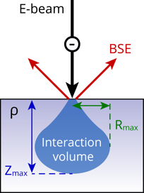
In order to answer this question, it is important to first understand how cathodoluminescence is generated. At room temperature, most electrons reside in the valence band, but when the electron beam excites the material, electrons move from valence band into a higher energy state (conduction band). After some characteristic time the electrons decay back into the valence band, and a photon can be emitted. If that happens, cathodoluminescence can be observed. The excitation of valence electrons by the electron beam occurs in a characteristic volume, which is normally referred to as the interaction volume.
Depending on whether a low voltage or high voltage electron beam interacts with a material, the interaction volume is either smaller or larger respectively. A higher energy electron beam penetrates deeper into the material, but because of the scattering in the material it also spreads more in the lateral direction leading to a reduction in the spatial resolution. Since electron have a high energy and can interact with the material more than once it is possible to excite multiple photons per primary electron. The higher the energy, the more photons can be generated typically.
For different materials, the interaction volume will vary for a given voltage due the different atomic mass and density. In order to help you assess which beam energy to select for an experiment, we created a database which can help you to find the “sweet spot” between high CL signal and high spatial resolution. This database consists of simulated electron trajectories in a range of materials (generated with CASINO, a Monte Carlo Simulation program of Electron Trajectories in Solids from the Université de Sherbrooke). There you can find electron-material interaction positions in 3D (X,Y,Z) and the associated electron energy (E) for such materials as Sapphire, Diamond, Zircon, Silicon, Gallium Nitride and Gallium Arsenide and more.
If you would like to know more about this, please register for the upcoming webinar session: Cathodoluminescence fundamentals: Imaging conditions for cathodoluminescence.
.png)


