.svg "DELMIC")
.png)


Perovskite materials have garnered significant attention from researchers and industry professionals, due to their exceptional photovoltaic and optoelectronic properties, as well as their low manufacturing cost. Among these materials, the exceptional optoelectronic properties of organic-inorganic hybrid perovskites (OIHPs) have led to rapid progress in improving the power conversion efficiency of photovoltaic cells. [1,2,3,4]
OIHPs have unique optoelectronic properties, such as a direct band gap, sharp absorption edge [5,6,7], large photocarrier diffusion lengths [8,9], and high tolerance to defects and grain boundaries [10,11]. Additionally, OIHPs exhibit tunability [13], moderate mobilities of charge carriers [8] and ions [16], strong polaronic coupling [12,15], and a prominent photostriction effect [16]. These properties make OIHPs versatile candidates for a wide array of optoelectronic applications beyond solar cells. However, the stability of OIHPs is a crucial limiting factor for long-term practical applications. Many extrinsic factors, such as moisture [18,19], oxygen [20], thermal decomposition [21], and light-induced chemical reactions [22,23], can lead to the degradation of these materials [17,18].
The challenge of ion migration in perovskites
The instability of OIHPs is related to their weak chemical bonds [24] and strongly anharmonic lattice dynamics [25], which lead to ultralow thermal conductivity [26] and facile ion migration induced by temperature [27], electric fields [28], or light exposure [29]. Ion migration is an intrinsic process in OIHPs [30,31] and has been observed to impact material morphology [28], composition, and device stability [23,30,32]. While prior studies have reported light-induced migration of different ion species in OIHPs [33], these ions' detailed microscopic migration pathways still need to be clarified [30].
A 2023 study published in Nature Communications focuses on investigating the photo-induced ion migration in OIHPs, specifically MAPbI3 and FAPbI3, using a combination of in situ scanning electron microscopy (SEM), energy-dispersive X-ray spectroscopy (EDS), and cathodoluminescence (CL) techniques.
The goal is to directly map the three-dimensional (3D) ion migration pathways, both laterally and vertically, and correlate this information with the optoelectronic properties of OIHPs. This is critical for designing more stable and efficient devices based on these materials.
The methodology behind the study
The researchers utilized in situ laser illumination inside a Scanning Electron Microscope (SEM), coupled with secondary electron imaging, Energy Dispersive Spectroscopy (EDS), and Cathodoluminescence (CL) with varying primary electron energies, to characterize the ion migration processes in perovskite materials. The study focused on two model systems: methylammonium lead iodide and formamidinium lead iodide.
Preparing the perovskite samples
The fabrication process for the perovskite samples began with the meticulous cleaning of FTO-coated glass substrates, followed by the preparation of two different perovskite precursor solutions - a 1.5M MAPbI3 precursor solution and a 1.4M FAPbI3 precursor solution.
The MAPbI3 precursor solution was made by dissolving equal molar ratios of methylammonium iodide (MAI) and lead iodide (PbI2) in a solvent mixture of dimethylformamide (DMF) and dimethyl sulfoxide (DMSO) with a volume ratio of 4:1. Similarly, the FAPbI3 precursor solution was prepared by dissolving equal molar ratios of formamidinium iodide (FAI) and PbI2 in a DMF and DMSO solvent mixture with a volume ratio of 8:1.
The perovskite precursor solutions were then spin-coated onto the cleaned FTO substrates at 4000 rpm for 30 seconds. During the spin-coating process, 0.6 ml of diethyl ether was dripped onto the substrate in the last 15 seconds. The as-spun films were subsequently annealed, with the MAPbI3 films annealed at 100°C for 10 minutes and the FAPbI3 films annealed at 150°C for 20 minutes.
Quantifying ion migration using SEM and EDS measurement
The study leveraged the power of SEM and EDS to capture secondary electron images and analyze elemental compositions. The SEM and EDS measurements in this research were performed using a FEG-SEM (Field Emission Gun – Scanning Electron Microscope) at the University of California Santa Barbara. The secondary electron imaging was detected using a standard Everhart-Thornley detector. At the same time, the EDS spectra were collected using an Energy Dispersive Spectroscopy (EDS) detector and then interpreted. Throughout the measurements, a high vacuum of 1 × 10⁻⁶ torr was maintained inside the SEM chamber to ensure the accuracy of the results.
For the light exposure tests, a Yb-doped fiber laser (Clark-MXR IMPULSE) with a fundamental wavelength of 1030 nm, an average pulse width of 150 fs, and a repetition rate of 5 MHz was utilized. The fundamental wavelength was converted to 515 nm using a BBO crystal and conveyed into the SEM chamber through a transparent viewport on the chamber wall. Each light exposure test was conducted on a fresh area of the sample to ensure the reliability of the results.
Following light exposure, a secondary electron image of the exposed area was captured with an acceleration voltage of 5 keV. A low beam current of 300 pA was used to minimize electron-beam-induced damage to the sample. A dwell time of 1 μs per pixel was used for imaging. Additionally, EDS line scans were performed across the center of the light-exposed area at approximately 7 μm intervals, with a beam current of 300 pA. To enhance the signal-to-noise ratio and minimize electron-beam-induced damage, 100 linescans, each taking 100 ms per scan, were captured and averaged.
Furthermore, the change in the absolute count of X-ray photons emitted from the exposed area was calculated and compared to the pristine background area. This allowed for the quantification of the impact of light exposure on the sample.
Key findings from the EDS measurement
1. Iodine ions are the most mobile species in OIHPs. EDS analysis shows a reduction in iodine X-ray counts in the illuminated region, indicating a deficiency of iodine due to the formation of I2 vapor that escapes the sample. The spatial profile of the iodine deficiency expands over time, suggesting iodine ion migration over a length scale of hundreds of micrometers, driven by the iodine concentration gradient. The estimated iodine ion diffusivity is around 3.5 × 10^-10 cm^2/s, significantly higher than that measured in single-crystal perovskites.
2. Lead ions primarily migrate from the bulk towards the sample surface. While the lead distribution probed by high-energy electrons (30 keV) remained unchanged, the lead distribution near the surface probed by low-energy electrons (5 keV) showed a significant increase and a double-peak structure after prolonged light exposure, corresponding to the bright ring contrast observed in secondary electron images.
3. The X-ray counts associated with silicon and tin significantly increased with light exposure, as probed by the high-energy electrons. This suggests the migration of silicon and tin ions, possibly induced by lead vacancies created in the bulk of the FAPbI3 sample.
4. The organic ions (nitrogen, carbon, and oxygen) [34] also showed a decrease in concentration near the sample surface due to light exposure. Still, their diffusivity could not be quantified due to low EDS sensitivity.
EDS elemental analysis of FAPbI3 after optical exposure

(A) X-ray count changes for iodine, detected by 30-keV photoelectrons (PEs), relative to unexposed areas. (B) Estimated iodine depletion radii versus exposure time, analyzed by 30-keV PEs, with error bars showing 95% confidence intervals. Additionally, EDS linescans for: (C) Lead (30-keV PEs), (D) Lead (5-keV PEs), (E) Tin (30-keV PEs), and (F) Silicon (30-keV PEs). All counts normalized to pre-exposure values.
Photophysical exploration of perovskites using cathodoluminescence
Barbé et al. [37] observed a ring-shaped area with enhanced photoluminescence in MAPbI3, which they attributed to the formation of a thin layer of PbI2. This observation was made using Raman spectroscopy. However, they did not find direct evidence of photoluminescence near the bandgap of PbI2. This was due to the use of a 532-nm excitation laser, which may not effectively excite PbI2, especially at ambient temperatures. So the researchers turned to cathodoluminescence (CL) with in situ laser exposure to effectively probe spatial heterogeneities in perovskite excitonic and defect emission.
The CL measurements were performed using a Delmic SPARC CL collection module integrated into an Environmental scanning electron microscope (ESEM) at the Center for Nanophase Materials Sciences (CNMS) at the Oak Ridge National Laboratory.
The experimental setup involved the use of a parabolic mirror to collect the CL generated under electron-beam excitation and to deliver the laser to the sample. A retractable mirror was used to direct free-space coupled laser sources, which included a 495-nm pulsed laser source and a 532-nm continuous-wave (CW) laser source, to the parabolic mirror. The 495-nm pulsed laser had a pulse duration of 100 fs and a repetition rate of 80 MHz, and it was focused to a spot size of 5 μm on the sample.
The optical penetration depth of the 495-nm laser in both MAPbI3 and FAPbI3 was approximately 70 nm [3]. After variable laser exposure times with the retractable mirror inserted, the mirror was retracted, and CL spectrum images were acquired using the electron-beam conditions described in the manuscript. To compensate for any movement of the SEM relative to the optics table, an active beam stabilization system was used.
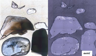
Cathodoluminescence analysis of MAPbI3 film after in situ laser exposure

Scanning electron microscopy (SEM) images (first column) and cathodoluminescence (CL) decomposition (columns 2–5) after exposure to a 495-nm, 100-μW fs laser with a 5-μm spot size for 90–600 seconds. Non-negative matrix factorization (NMF) identifies three CL components: MAPbI3 exciton (red), partially degraded MAPbI3 (blue), and PbI2 (purple). The PbI2 CL signal (final column), integrated from 500-520 nm, is much weaker. Scale bar: 5 μm.
The researchers employed blind non-negative matrix factorization (NMF) to analyze the CL spectrum images and track changes in the CL spectra as a function of laser exposure time. This approach allowed them to investigate the photophysical properties and dynamics of the hybrid perovskite materials under laser illumination.
Key findings from cathodoluminescence
1. CL microscopy was used to probe spatial heterogeneities in perovskite excitonic and defect emission. The CL analysis indicated complete suppression of PbI2 emission within the laser-exposed region, suggesting that the ion migration induced by laser exposure did not introduce any near-surface PbI2 or partially decomposed phases.
2. The secondary electron (SE) images showed increased brightness and morphological changes after laser exposure, consistent with previous reports. The size of the microstructured area increased with increasing laser exposure time.
3. The CL non-negative matrix factorization (NMF) decomposition revealed that component 1 was consistent with excitonic luminescence, while component 2 was consistent with near-band-edge CL from an intermediate phase resulting from partial degradation of the film [34,35]. The raw PbI2 CL showed that the PbI2 CL was completely suppressed in the area around the laser spot, and the area with suppressed PbI2 CL increased with increasing laser exposure time.
4. At higher laser powers, thermal effects started to emerge, and the excitonic CL was also suppressed. However, the hybrid perovskite films exhibited substantially enhanced luminescence intensity in the area surrounding the laser spot under these conditions.
5. The authors concluded that the observed effects were due to photo-induced ion migration rather than thermal decomposition, as evidenced by the long-range changes in SE contrast, the changes in absolute X-ray counts of lead, and the lack of spectral evidence for the thermal decomposition product PbI2.
Conclusion
The study provides important insights into the ion migration processes in perovskites, which can aid in the design and processing of perovskite materials for future applications. Understanding the ion migration pathways is crucial for addressing the challenges of material instability and photocurrent hysteresis in perovskite solar cells, which have hindered their practical implementation.
This article is based on the study - https://www.nature.com/articles/s41467-023-37486-w
References
- Jung, E. H., Jeon, N. J., Park, E. Y., Moon, C. S., Shin, T. J., Yang, T., Noh, J. H., & Seo, J. (2019). Efficient, stable and scalable perovskite solar cells using poly(3-hexylthiophene). Nature, 567(7749), 511–515. https://doi.org/10.1038/s41586-019-1036-3
- Jiang, Q., Tong, J., Xian, Y., Kerner, R. A., Dunfield, S. P., Xiao, C., Scheidt, R. A., Kuciauskas, D., Wang, X., Hautzinger, M. P., Tirawat, R., Beard, M. C., Fenning, D. P., Berry, J. J., Larson, B. W., Yan, Y., & Zhu, K. (2022). Surface reaction for efficient and stable inverted perovskite solar cells. Nature, 611(7935), 278–283. https://doi.org/10.1038/s41586-022-05268-x
- Kim, J. Y., Lee, J., Jung, H. S., Shin, H., & Park, N. (2020). High-Efficiency Perovskite solar cells. Chemical Reviews, 120(15), 7867–7918. https://doi.org/10.1021/acs.chemrev.0c00107
- Park, S. Y., & Zhu, K. (2022). Advances in SnO2 for Efficient and Stable n–i–p Perovskite Solar Cells. Advanced Materials, 34(27). https://doi.org/10.1002/adma.202110438
- Baikie, T., Fang, Y., Kadro, J. M., Schreyer, M., Wei, F., Mhaisalkar, S. G., Graetzel, M., & White, T. J. (2013). Synthesis and crystal chemistry of the hybrid perovskite (CH3NH3)PbI3 for solid-state sensitised solar cell applications. Journal of Materials Chemistry. A, 1(18), 5628. https://doi.org/10.1039/c3ta10518k
- Stranks, S. D., & Snaith, H. J. (2015). Metal-halide perovskites for photovoltaic and light-emitting devices. Nature Nanotechnology, 10(5), 391–402. https://doi.org/10.1038/nnano.2015.90
- Yin, W., Shi, T., & Yan, Y. (2015). Superior Photovoltaic Properties of Lead Halide Perovskites: Insights from First-Principles Theory. Journal of Physical Chemistry. C./Journal of Physical Chemistry. C, 119(10), 5253–5264. https://doi.org/10.1021/jp512077m
- Shi, D., Adinolfi, V., Comin, R., Yuan, M., Alarousu, E., Buin, A., Chen, Y., Hoogland, S., Rothenberger, A., Katsiev, K., Losovyj, Y., Zhang, X., Dowben, P. A., Mohammed, O. F., Sargent, E. H., & Bakr, O. M. (2015). Low trap-state density and long carrier diffusion in organolead trihalide perovskite single crystals. Science, 347(6221), 519–522. https://doi.org/10.1126/science.aaa2725
- Guo, Z., Wan, Y., Yang, M., Snaider, J., Zhu, K., & Huang, L. (2017). Long-range hot-carrier transport in hybrid perovskites visualized by ultrafast microscopy. Science, 356(6333), 59–62. https://doi.org/10.1126/science.aam7744
- Ball, J. M., & Petrozza, A. (2016). Defects in perovskite-halides and their effects in solar cells. Nature Energy, 1(11). https://doi.org/10.1038/nenergy.2016.149
- Brenes, R., Guo, D., Osherov, A., Noel, N. K., Eames, C., Hutter, E. M., Pathak, S. K., Niroui, F., Friend, R. H., Islam, M. S., Snaith, H. J., Bulović, V., Savenije, T. J., & Stranks, S. D. (2017). Metal halide perovskite polycrystalline films exhibiting properties of single crystals. Joule, 1(1), 155–167. https://doi.org/10.1016/j.joule.2017.08.006
- Grancini, G., & Nazeeruddin, M. K. (2018). Dimensional tailoring of hybrid perovskites for photovoltaics. Nature Reviews. Materials, 4(1), 4–22. https://doi.org/10.1038/s41578-018-0065-0
- Kulkarni, S. A., Baikie, T., Boix, P. P., Yantara, N., Mathews, N., & Mhaisalkar, S. (2014). Band-gap tuning of lead halide perovskites using a sequential deposition process. Journal of Materials Chemistry. A, 2(24), 9221–9225. https://doi.org/10.1039/c4ta00435c
- Bakulin, A. A., Selig, O., Bakker, H. J., Rezus, Y. L., Müller, C., Glaser, T., Lovrincic, R., Sun, Z., Chen, Z., Walsh, A., Frost, J. M., & Jansen, T. L. C. (2015). Real-Time observation of organic cation reorientation in methylammonium lead iodide perovskites. the Journal of Physical Chemistry Letters, 6(18), 3663–3669. https://doi.org/10.1021/acs.jpclett.5b01555
- Kandada, A. R. S., & Silva, C. (2020). Exciton polarons in Two-Dimensional hybrid Metal-Halide Perovskites. Journal of Physical Chemistry Letters, 11(9), 3173–3184. https://doi.org/10.1021/acs.jpclett.9b02342
- Zhou, Y., You, L., Wang, S., Ku, Z., Fan, H., Schmidt, D., Rusydi, A., Chang, L., Wang, L., Ren, P., Chen, L., Yuan, G., Chen, L., & Wang, J. (2016). Giant photostriction in organic–inorganic lead halide perovskites. Nature Communications, 7(1). https://doi.org/10.1038/ncomms11193
- Boyd, C. C., Cheacharoen, R., Leijtens, T., & McGehee, M. D. (2018). Understanding degradation mechanisms and improving stability of perovskite photovoltaics. Chemical Reviews, 119(5), 3418–3451. https://doi.org/10.1021/acs.chemrev.8b00336
- Bella, F., Griffini, G., Correa-Baena, J., Saracco, G., Grätzel, M., Hagfeldt, A., Turri, S., & Gerbaldi, C. (2016). Improving efficiency and stability of perovskite solar cells with photocurable fluoropolymers. Science, 354(6309), 203–206. https://doi.org/10.1126/science.aah4046
- Christians, J. A., Herrera, P. a. M., & Kamat, P. V. (2015). Transformation of the Excited State and Photovoltaic Efficiency of CH3NH3PbI3 Perovskite upon Controlled Exposure to Humidified Air. Journal of the American Chemical Society, 137(4), 1530–1538. https://doi.org/10.1021/ja511132a
- Bryant, D., Aristidou, N., Pont, S., Sanchez-Molina, I., Chotchunangatchaval, T., Wheeler, S., Durrant, J. R., & Haque, S. A. (2016). Light and oxygen induced degradation limits the operational stability of methylammonium lead triiodide perovskite solar cells. Energy & Environmental Science, 9(5), 1655–1660. https://doi.org/10.1039/c6ee00409a
- Conings, B., Drijkoningen, J., Gauquelin, N., Babayigit, A., D’Haen, J., D’Olieslaeger, L., Ethirajan, A., Verbeeck, J., Manca, J., Mosconi, E., De Angelis, F., & Boyen, H. (2015). Intrinsic thermal instability of methylammonium lead trihalide perovskite. Advanced Energy Materials, 5(15). https://doi.org/10.1002/aenm.201500477
- Domanski, K., Roose, B., Matsui, T., Saliba, M., Turren-Cruz, S., Correa-Baena, J., Carmona, C. R., Richardson, G., Foster, J. M., De Angelis, F., Ball, J. M., Petrozza, A., Mine, N., Nazeeruddin, M. K., Tress, W., Grätzel, M., Steiner, U., Hagfeldt, A., & Abate, A. (2017). Migration of cations induces reversible performance losses over day/night cycling in perovskite solar cells. Energy & Environmental Science, 10(2), 604–613. https://doi.org/10.1039/c6ee03352k
- Burschka, J., Pellet, N., Moon, S., Humphry-Baker, R., Gao, P., Nazeeruddin, M. K., & Grätzel, M. (2013). Sequential deposition as a route to high-performance perovskite-sensitized solar cells. Nature, 499(7458), 316–319. https://doi.org/10.1038/nature12340
- Ma, H., Li, C., Ma, Y., Wang, H., Rouse, Z. W., Zhang, Z., Slebodnick, C., Alatas, A., Baker, S. P., Urban, J. J., & Tian, Z. (2019). Supercompliant and soft (CH3NH3)3Bi. Physical Review Letters, 123(15). https://doi.org/10.1103/physrevlett.123.155901
- Yaffe, O., Guo, Y., Tan, L. Z., Egger, D. A., Hull, T., Stoumpos, C. C., Zheng, F., Heinz, T. F., Kronik, L., Kanatzidis, M. G., Owen, J. S., Rappe, A. M., Pimenta, M. A., & Brus, L. E. (2017). Local polar fluctuations in lead halide perovskite crystals. Physical Review Letters, 118(13). https://doi.org/10.1103/physrevlett.118.136001
- Haque, M. A., Kee, S., Villalva, D. R., Ong, W., & Baran, D. (2020). Halide perovskites: Thermal transport and prospects for thermoelectricity. Advanced Science, 7(10). https://doi.org/10.1002/advs.201903389
- Lai, M., Obliger, A., Lu, D., Kley, C. S., Bischak, C. G., Kong, Q., Lei, T., Dou, L., Ginsberg, N. S., Limmer, D. T., & Yang, P. (2018). Intrinsic anion diffusivity in lead halide perovskites is facilitated by a soft lattice. Proceedings of the National Academy of Sciences of the United States of America, 115(47), 11929–11934. https://doi.org/10.1073/pnas.1812718115
- Xiao, Z., Yuan, Y., Shao, Y., Wang, Q., Dong, Q., Bi, C., Sharma, P., Gruverman, A., & Huang, J. (2014). Giant switchable photovoltaic effect in organometal trihalide perovskite devices. Nature Materials, 14(2), 193–198. https://doi.org/10.1038/nmat4150
- Kim, G. Y., Senocrate, A., Yang, T., Gregori, G., Grätzel, M., & Maier, J. (2018). Large tunable photoeffect on ion conduction in halide perovskites and implications for photodecomposition. Nature Materials, 17(5), 445–449. https://doi.org/10.1038/s41563-018-0038-0
- Yuan, Y., & Huang, J. (2016). Ion migration in organometal trihalide perovskite and its impact on photovoltaic efficiency and stability. Accounts of Chemical Research, 49(2), 286–293. https://doi.org/10.1021/acs.accounts.5b00420
- Bi, E., Song, Z., Li, C., Wu, Z., & Yan, Y. (2021). Mitigating ion migration in perovskite solar cells. Trends in Chemistry, 3(7), 575–588. https://doi.org/10.1016/j.trechm.2021.04.004
- Snaith, H. J., Abate, A., Ball, J. M., Eperon, G. E., Leijtens, T., Noel, N. K., Stranks, S. D., Wang, J. T., Wojciechowski, K., & Zhang, W. (2014). Anomalous hysteresis in perovskite solar cells. Journal of Physical Chemistry Letters, 5(9), 1511–1515. https://doi.org/10.1021/jz500113x
- Deng, Y., Xiao, Z., & Huang, J. (2015). Light‐Induced Self‐Poling effect on organometal trihalide perovskite solar cells for increased device efficiency and stability. Advanced Energy Materials, 5(20). https://doi.org/10.1002/aenm.201500721
- Scanning electron microscopy and X-Ray microanalysis. (n.d.). SpringerLink. https://link.springer.com/book/10.1007/978-1-4615-0215-9
- Xiao, C., Li, Z., Guthrey, H., Moseley, J., Yang, Y., Wozny, S., Moutinho, H., To, B., Berry, J. J., Gorman, B., Yan, Y., Zhu, K., & Al-Jassim, M. (2015). Mechanisms of Electron-Beam-Induced damage in perovskite thin films revealed by cathodoluminescence spectroscopy. Journal of Physical Chemistry. C./Journal of Physical Chemistry. C, 119(48), 26904–26911. https://doi.org/10.1021/acs.jpcc.5b09698
- Taylor, E. J., Iyer, V., Dhami, B. S., Klein, C., Lawrie, B. J., & Appavoo, K. (2022, January 17). Hyperspectral nanoscale mapping of hybrid perovskite photophysics at the single grain level. arXiv.org. https://arxiv.org/abs/2201.06546
- Barbé, J., Newman, M., Lilliu, S., Kumar, V., Lee, H. K. H., Charbonneau, C., Rodenburg, C., Lidzey, D., & Tsoi, W. C. (2018). Localized effect of PbI2 excess in perovskite solar cells probed by high-resolution chemical–optoelectronic mapping. Journal of Materials Chemistry. A, 6(45), 23010–23018. https://doi.org/10.1039/c8ta09536a