.svg "DELMIC")
.png)


Light, being an electromagnetic wave, has an amplitude, wavelength, wave vector, and polarisation. All of these properties of the emitted CL, in turn, provide information about different sample properties and can be studied using a range of imaging modes such as intensity mapping, spectroscopy, angle-resolved imaging, and polarimetry.
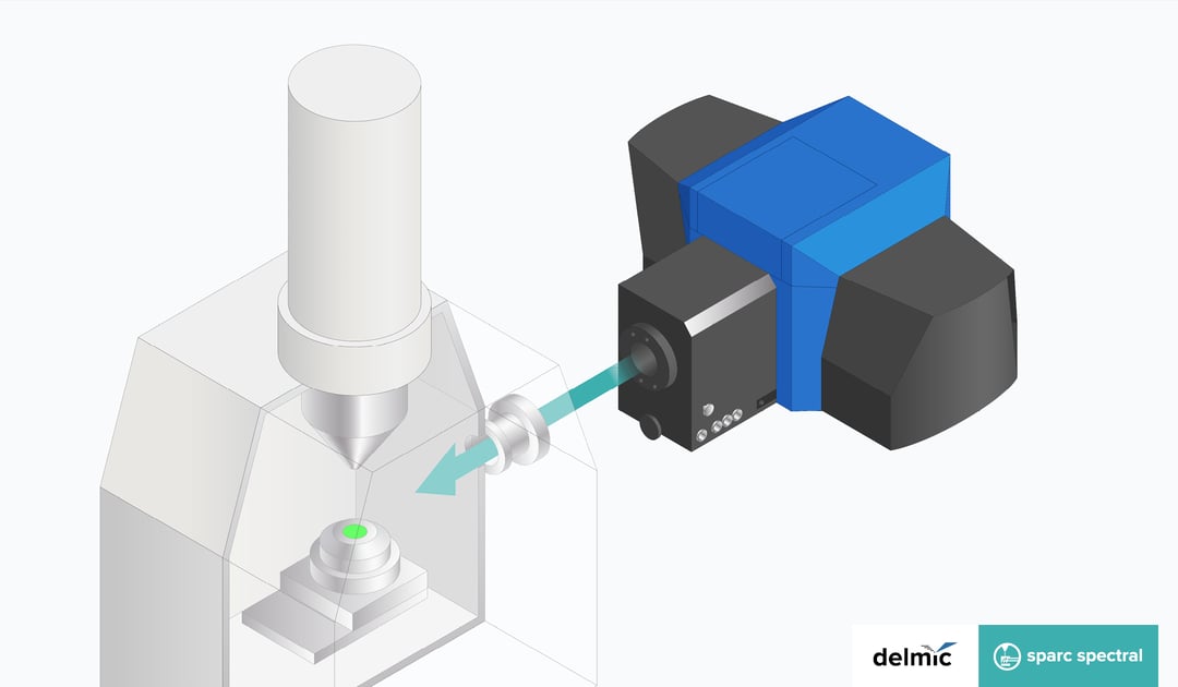
Cathodoluminescence imaging is often performed in a Scanning Electron Microscope (SEM). One of the main benefits is the focussed electron beam - spot sizes of a few nanometres are typical, which can be used to excite the sample very locally. While conventional optical techniques are diffraction-limited, CL imaging in the SEM is a high-resolution optical technique. Moreover, the energy of the electron beam in the SEM can be tuned, therefore affording the possibility of tuning the excitation. Some samples are sensitive to electron exposure and cannot withstand high electron energies, whereas some samples, such as plasmonic nanostructures, require high electron energy to be excited. Some materials, such as plastics, exhibit charging at high current, whereas some have low CL emission, requiring the use of high excitation currents. Performing CL imaging in the SEM allows us to optimize the excitation parameters to get the maximum signal from the sample while ensuring minimum or no beam damage.
The SEM is also a great platform for performing correlative imaging with CL, together with techniques like EDX (Energy Dispersive X-ray spectroscopy), SE (Secondary Electron), BSE (Backscattered Electron), and EBSD (Electron Backscattered Diffraction). All of these imaging techniques provide different and often complementary types of information about the sample: SE imaging provides topographic contrast, whereas BSE imaging provides primarily material contrast (from material density). EDX is based on core transitions in the material and provides quantitative information on the material composition, and EBSD allows us to probe the crystal structure and orientation.
The CL contrast can provide information on crystal defects, ionization state, and composition. SEM-based correlative CL imaging is a powerful and versatile technique by which a wide range of material properties can be studied, and is being used in fields as diverse as semiconductors, solar cells, plasmonics, geology, life sciences, and materials sciences.
.gif?width=227&name=CL%20FAQ_Animated%20Banner%20(1).gif)


