.svg "DELMIC")
.png)


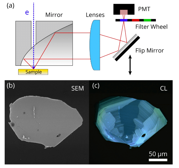
Various geological samples, including minerals, rocks and crystals, can be effectively imaged with intensity mapping. One of such examples is zircon crystal, a robust material, which is very resilient to weathering processes and can persist for a long time in a large variety of sedimentary, igneous, and metamorphic rocks. When using an RGB cathodoluminescence image, it is possible to visualise not only the contour of the zircon grain, but the structure within the grain. This information can be extremely valuable if you want to learn about the zircon growth conditions.
Apart from geological applications, CL intensity mapping can be used in other fields, such as materials science, to obtain images of dielectric, ceramic, semiconductor materials, as well as photovoltaic materials.
If you are interested in reading more about hyperspectral cathodoluminescence imaging, make sure to download the technical note below.