.svg "DELMIC")
.png)


There is a lot of interest in the field of polymers and plastics due to their importance in environmental studies. Raman spectroscopy is currently one of the main techniques used to characterize these materials. While this yields valuable information, it is a slow and expensive technique. Moreover, there are serious limitations where particle size is concerned and it is very challenging to characterize sub-micron particles. Therefore, a different technique is needed for the nanometer range. Delmic has performed CL imaging in collaboration with one of the labs of the European Commission to characterize different polymers and plastics in a range of particle sizes.
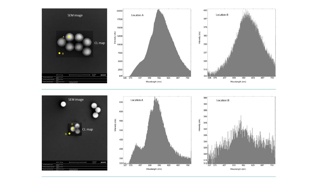
SEM-based CL imaging has the potential to be used as a characterization technique for these materials because of its resolution. It can be used in combination with SEM to rapidly locate and image large numbers of particles at high resolution. Using polystyrene as a test case, Delmic and our partner lab performed CL measurements on beads of various sizes, to determine if CL emission could be observed and whether the spectrum could be used to characterize the particles. Firstly, SEM imaging was performed to locate the beads, followed by the acquisition of a CL spectral map of the same region. The automated overlay of the CL and SEM images enables the characterization of the polymer material as you can see from the spectrum of location A in the image above. Detailed studies can be performed, for example, by studying the change in spectrum with particle size. Location B presents the background spectrum at a point outside the beads. The peaks can be distinguished very clearly over the background and we saw stable CL mission without any substantial signal lost. It should be noted that the background spectrum would be still weaker if location B were farther away from the beads. For imaging smaller particles such as 600 nm beads, we used a lower energy of 5 keV, because the high energy electron beam penetrates the bead and the substrate, resulting in CL emission not only from the bead but from the interaction volume. So a lower energy is better for imaging smaller particles . Once again, we observed stable CL mission and the spectral peaks could be clearly identified for the smaller polystyrene particles as well.
We have also applied this technique to other polymers and plastics like polyethylene and polypropylene, teflon, nylon, as well as mixtures of these materials. The SEM imaging parameters, like the energy and the current, and CL imaging conditions are currently being optimized for imaging particles of different sizes and materials. While the larger particles are easier to characterize because of the strong CL emission, the small particles which are less than 1 µm can be studied as well by performing a longer acquisition or background subtraction or both. CL imaging is therefore a very promising technique for the identification and characterization of polymer materials and micro plastics, and we are excited about the possibilities in this field.