.svg "DELMIC")
.png)


In nanophotonics, the strong interaction between light and matter taking place at the nanoscale is exploited to manipulate the flow and properties of light in carefully designed nanostructures. An important property in nanophotonic research is the radiation pattern, which describes the angular profile under which light is emitted from or scattered by a nanostructure. Nanoantennas, for example, can be designed to efficiently redirect light from or towards nanometer-sized point sources or collectors. Likewise, nanostructuring can be used to increase the outcoupling of light from light-emitting diodes (LEDs) or trap light in solar cells to increase their efficiency. Complex metasurfaces even hold the promise of creating ultrathin tunable lenses and on-chip lasers. [1] In this blog post, we will describe how cathodoluminescence (CL) imaging can help in understanding the complex radiation patterns encountered in nanophotonic research.
In angle-resolved cathodoluminescence imaging, the focused electron beam of a scanning electron microscope (SEM) is used as a broadband excitation source to excite structures with a nanometer spatial resolution. The interaction of the electron beam with the sample generates cathodoluminescence that is subsequently collected by a parabolic mirror and projected onto a 2D imaging array. Since every pixel in this image corresponds to a specific angle of emission, the radiation pattern of the investigated nanostructure can be reconstructed as a function of azimuthal φ and zenithal θ emission angles. The SPARC Spectral system uses a parabolic mirror with a high collection efficiency (89% for a Lambertian emitter) and a large acceptance angle (solid angle of 1.49π sr) to allow a wide range of emission angles to be measured in a single acquisition. Color filters can moreover be inserted in the beam path to investigate the angular profile of CL for different energy ranges.
The angular radiation patterns of nanostructures are strongly dependent on geometrical considerations such as their shape, local environment, and point of excitation. Owing to the small size of the structures involved, these effects are however difficult to investigate in detail using conventional diffraction-limited optical microscopy. Because angle-resolved CL imaging uses a focused electron beam instead of light as the excitation source, this technique can be used to study optical modes of nanostructures with a deep subwavelength resolution. The combination of high spatial resolution and angular resolving power offered by angle-resolved CL imaging allows for unprecedented characterization capabilities of nanophotonic structures. Not only does this open up new fundamental science [2], but it is also of crucial importance to establish design rules for nanophotonic applications.
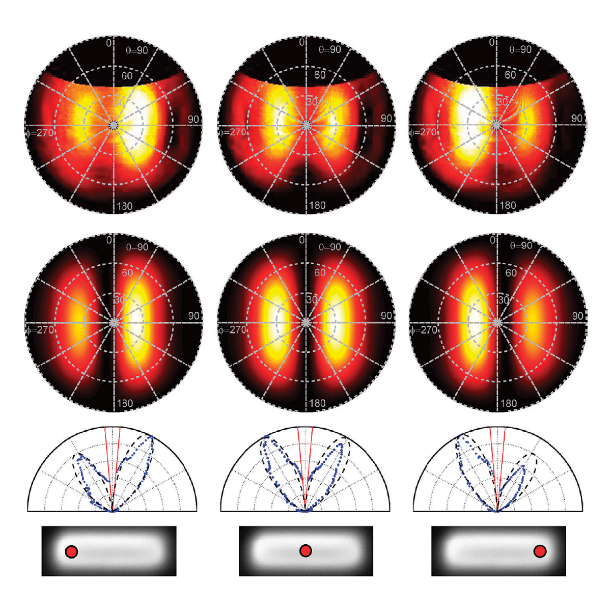
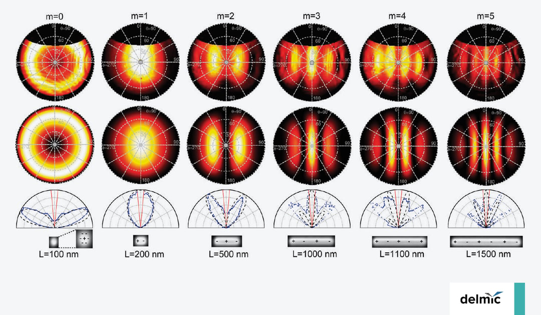
Gold nanorods are excited by the electron beam at different points along their length (top row), giving rise to the distinct radiation patterns below. Experimental angular profiles show good qualitative agreement with theory. Image adapted from ref [3].
An illustrative case of how angle-resolved CL imaging can be used to obtain detailed information on the light directing properties of nanophotonic structures is provided in the figure above. Gold ridge nanoantennas support plasmon resonance modes that can be excited directly by the electron beam. [3] Here, a 500 nm long gold plasmonic ridge nanoantenna is excited by the electron beam at different points along the antenna, as shown in the top row. The (simulated) polar plots of the angle-resolved CL show the radiation pattern resulting from such an oscillation, clearly showing how in this particular antenna light is radiated out preferentially along the long axis at an angle of approximately 30 degrees away from the surface normal. Using the high spatial resolution provided by the electron beam, it is possible to excite the nanoantenna close to their endpoints, leading to strong beaming of light towards the opposite end of the antenna. Experiments like these provide valuable information on how light from a point emitter, such as a quantum dot or nanodiamond can be redirected when positioned in close proximity to such a nanoantenna.
Angle-resolved CL imaging is a valuable experimental technique that can be used to study complex angular radiation profiles of nanophotonic structures with high spatial and angular resolution, providing deep insights into the light-matter interactions at the nanoscale. If you are interested to learn more about the application of cathodoluminescence imaging to nanophotonic research we recommend joining our upcoming webinar Merging metasurfaces with free electrons in the scanning electron microscope and visiting our dedicated webpage on plasmonics and metamaterials.
References
[1] A.F. Koenderink, A. Alù, and A. Polman, Science 348, 516 (2015)
[2] A. Polman, M. Kociak, and F.J. Garcia de Abajo, Nature Mater. 18, 1158 (2019)
[3] T. Coenen, E.J.R. Vesseur, and A. Polman, ACS Nano 6, 1742 (2012)