- What are the most common SEM techniques for studying materials?
- What kind of data can you get with these techniques?
- What makes cathodoluminescence (CL) different from other SEM techniques?
- 5 advantages of CL imaging
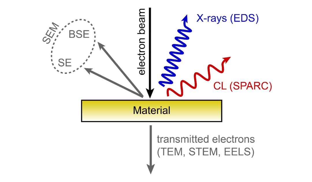
When studying or working with materials, it is essential to understand as much as possible about them. Electron microscopy techniques are a common way to get data, such as material contrast, composition, surface topography, and structure. When an electron beam (for example, in a scanning electron microscope) interacts with a material (bulk, thick or thin), multiple processes occur. Such interaction can generate secondary electrons (SE), backscattered electrons (BSE), and X-rays. Various techniques exist that harness these signals. The most commonly used techniques are secondary electron (SE) and backscattered electron (BSE) detection, electron backscatter diffraction (EBSD), and energy-dispersive X-ray imaging (EDS), which can be used to obtain various types of information about the material.
Secondary electrons (SE) detection is the detection of low-energy electrons, with which it is possible to collect secondary electrons only from the top few nanometers of the material. This technique is sensitive to surface topography and also shows (minor) material contrast.
Backscattered electrons (BSE) detection is primarily sensitive to density and atomic number and as such can be used to obtain material contrast.
With electron backscattered diffraction (EBSD) you can look at the crystal structure and crystal orientation.
Energy-dispersive X-ray spectroscopy (EDS) probes core transitions in the material and as such can be used for quantitative elemental analysis
Next to the above-mentioned signals, light can also be generated during the electron beam interaction with a material. This is known as cathodoluminescence (CL).
What makes cathodoluminescence different from other SEM techniques?
Cathodoluminescence provides unique and complementary information to the other SEM-based techniques. First of all, it allows observing an emission energy range of 0.5 to 6 eV. In this energy range information about the composition, crystal structure, and the electronic band gap can be obtained for example. Furthermore, trace elements or dopants can be sensitively detected with CL because they have different transitions than bulk materials. Similarly, it is possible to look at crystal defects as these can alter the local optical properties of the material. With CL you can image optical resonances in a range of (resonant) photonic systems and you are sensitive to the refractive index of the materials. Overall, it is a powerful tool and a visible measure of what is taking place inside a material or an organism. Combined with other SEM-based techniques, it can be used to produce the most complete material analysis.
-
Nanoscale excitation resolution well below the optical diffraction limit
-
Probeless and contactless inspection technique (compared to near-field microscopy, for example, a physical probe is not required);
-
Broadband excitation allows inspection from the deep UV to the infrared spectral range;
-
The spectral resolution is high in state-of-the-art CL systems allowing high-quality spectroscopy;
-
The electron penetration depth is tunable, which allows depth-resolved studies and imaging of buried structures
If you are looking for more information explaining the basics of cathodoluminescence, make sure to check out the webinar series called Cathodoluminescence Fundamentals. Over the course of six webinars, we discuss the basics of cathodoluminescence, from CL processes to sample preparation and application in geology, semiconductors, and life sciences.
.png)


