.svg "DELMIC")
.png)


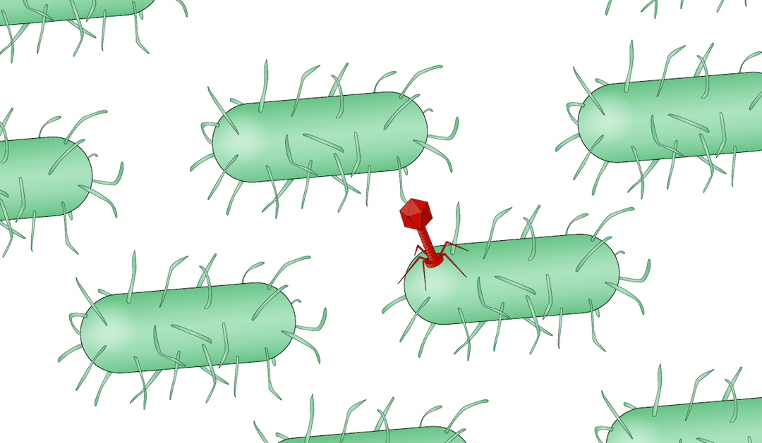
Viruses have long been studied using electron microscopy (EM) to understand their mechanisms of pathogenesis. Typically, viral particles are isolated and purified, after which they are imaged in an EM to determine the structure of the virus. Unfortunately, this approach fails to provide insight into the interaction between the virus and host cells. Cryogenic electron tomography (cryo-ET) allows the study of viruses in their near-native cellular environment and is thus able to reveal the interaction between the virus and its host. Most samples will require thinning steps to get a high-resolution tomogram. The most artefact free way to thin your sample is focused ion beam (FIB) milling: using this method you can create a thin section of your cells called a lamella.
Viral infection studied in situ with cryo-ET
Although cryo-ET is a relatively new method, the first examples available show the power of the technique in viral infection research. The combination of Cryo-ET and FIB milling has been used to examine the replication process of bacteriophages within different bacteria in detail. Using cryo-ET, many of the important steps in the assembly of the bacteriophages were then visualized, as well as the compartments formed to protect the genetic material of the bacteriophages within the bacteria [2-4]. Hagen et al. have also used a cryo-FIB and ET workflow to study the nuclear envelopment of herpesvirus capsids at high resolution [5].
Using FLM to guide FIB milling
Current publications using cryo-ET focus on general events during the infection because specific structures or transient events are hard to capture. To solve this problem fluorescent light microscopy can be used to find the structure of interest and ensure this structure is present in a prepared lamella. Currently, this means that users have to transfer their sample to a separate cryogenic fluorescent light microscope, making the cryo-ET workflow more error-prone. Delmic has developed a fluorescent light microscope that can be retrofitted to your current FIB/SEMFocused-ion beam /scanning electron microscope called METEOR. This solution will simplify the workflow allowing researchers to study transient events during viral infection.
Bulk cryo-ET to study viral infection
Many viruses evolve very rapidly due to the high mutation rate and short replication time. This evolution is dangerous to human health, as it enables viruses to escape immunity and drugs. Cryo-ET could be used to examine the structural changes of the viruses during evolution. Additionally, cryo-ET could be employed for comparative studies. For example, viruses from the same viral family, mutants or infection intermediates could be compared.
Currently, doing these kinds of experiments would mean having to prepare a large number of grids and performing the laborious workflow many times. Delmic is developing an automated sample transfer system that holds up to 12 grids and is compatible with autoloader systems on most existing cryo-TEM systems. This results in a workflow that abolishes the need for manual handling of grids after sample vitrification and loading into the cassette. By employing this technique large screening experiments of viruses using cryo-ET become a possibility.
Cryo-ET is a powerful technique that enables researchers to understand viral infections better. An integrated fluorescent light microscope and automated sample transfer will allow researchers to gain even more information and deepen their understanding of viral infections.
References
[1] Lamers MM, Beumer J, van der Vaart J, et al. SARS-CoV-2 productively infects human gut enterocytes. Science. Published online May 1, 2020:eabc1669. doi:10.1126/science.abc1669
[2] Chaikeeratisak V, Nguyen K, Khanna K, et al. Assembly of a nucleus-like structure during viral replication in bacteria. Science. 2017;355(6321):194-197. doi:10.1126/science.aal2130
[3] Dai W, Fu C, Raytcheva D, et al. Visualizing virus assembly intermediates inside marine cyanobacteria. Nature. 2013;502(7473):707-710. doi:10.1038/nature12604
[4] Chaikeeratisak V, Khanna K, Nguyen KT, et al. Viral Capsid Trafficking along Treadmilling Tubulin Filaments in Bacteria. Cell. 2019;177(7):1771-1780.e12. doi:10.1016/j.cell.2019.05.032
[5] Hagen C, Dent KC, Zeev-Ben-Mordehai T, et al. Structural Basis of Vesicle Formation at the Inner Nuclear Membrane. Cell. 2015;163(7):1692-1701. doi:10.1016/j.cell.2015.11.029
This work is supported by the European SME2 grant № 879673 - Cryo-SECOM Workflow