.svg "DELMIC")
.png)


Fluorescence microscopy is considered a reliable tool for studying organic samples at a high resolution. Nevertheless, the diffraction barrier of light poses the problem of not being able to distinguish objects that are smaller than the wavelength of light. Proteins, for example, can be as small as four nanometers. Even recent developments in light microscopy - such as super-resolution which can resolve objects at a smaller scale - come with the problem of providing no contextual or structural information besides the objects that glow under fluorescence.
It is common practice to utilize multiple tools to obtain data of the same sample. Scientists might therefore combine their data obtained from a light microscope with that of a separate electron microscope. This, however, poses the challenge of unwanted changes that occur in the sample in between measurements. A high level of interpretation is also required of the scientist, who needs to manually overlay the images.
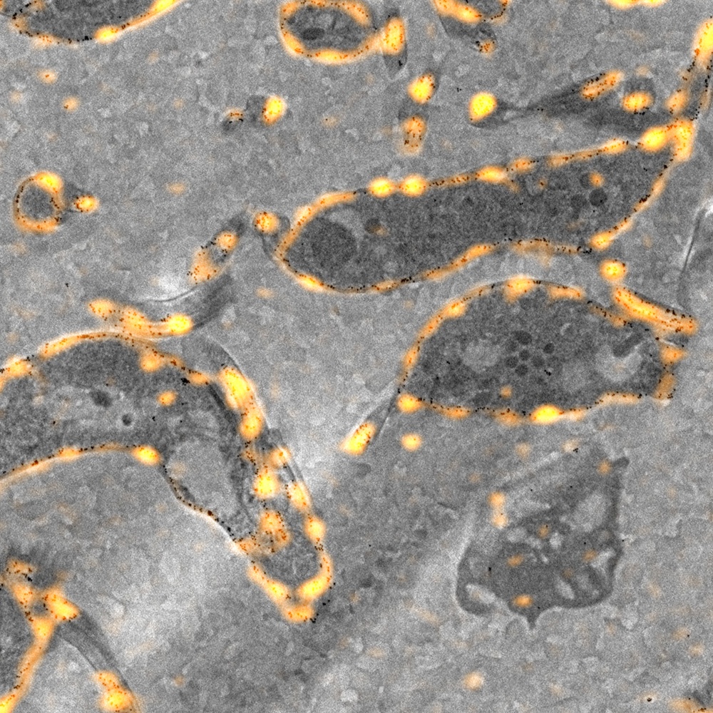
In an integrated system, data from the fluorescence and electron microscope are obtained simultaneously. This is possible with the right tools and methods: a fluorescence microscope which can be retrofitted on a scanning electron microscope, and the correct sample preparation methods which enables the sample to be measured under both a fluorescence and electron microscope.
Electron microscopy has great potential as a tool to supplement data which needs to viewed under a light microscope. EM can, for instance, illuminate the area surrounding your region of interest with the high-resolution data delivered by the electron beam.


