.svg "DELMIC")
.png)


Minerals and rocks can go through various deformation mechanisms, which are microscopic changes in the internal structure, shape, and volume of a material. Deformation mechanisms include processes such as [1]:
- Recrystallization
- Cataclasis
- Dislocation creep
- Diffusion creep
In both natural and experimental deformation and fracturing (faulting) processes, fluid-rock interactions often play a significant role [2].
One mechanism for deformation is called dissolution-precipitation creep (DPC), which involves the dissolution and precipitation of material during diffusion creep. Its presence is usually identified through signs of dissolution, such as irregular grain boundaries and reduced porosity [3]. However, it’s usually not known at which location the material has precipitated, as distinguishing precipitation from the original grains can be challenging.
Knowing the location of the precipitated material is crucial, as it provides valuable information about the role of solution transfer processes in deformation and the involved length scales in diffusive mass transfer. The latter factor ultimately determines the overall deformation rate produced by the processes of dissolution and precipitation [2].
Low-velocity shear experiments
Cathodoluminescence (CL) and hyperspectral imaging are powerful techniques for investigating the microscale variations in the structure of minerals. In a recent study by Hamers et al., researchers used CL imaging to investigate quartz precipitation during two low-velocity shear experiments [2]. One experiment used a pure quartz gouge, while the other used a mixed quartz and muscovite gouge. They chose these materials because their CL signal reveals their dissolution-precipitation microstructures.
To conduct the shear experiments, the researchers used a hydrothermal ring shear apparatus. They obtained false-color images using Delmic’s SPARC Compact CL system, among others. They also conducted hyperspectral CL imaging as part of their analysis.
Increased Al concentration
In both shear experiments, there was a clear distinction visible between the starting material and the deformed material. They observed microstructural evidence for dissolution-precipitation processes, as well as blue luminescent quartz between the cracks and pores of the deformed material, which was not present in the starting material (Figure 1).
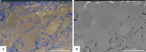 Figure 1 CL and BSE images of the sheared quartz gouge. a) False-colour RGB CL image overlay on BSE image of the pulverized zone next to the slip surface. Fine-grained red luminescent quartz fragments are sealed together by blue luminescent quartz in fractures, pore space, and grain rims. (b) BSE image corresponding to the CL image in (a). Ni-rich material, injected from the slip surface into the gouge, is recognized by the high brightness (dark in CL). Taken from [2].
The researchers concluded that during the experiments, precipitation of quartz with increased Al concentration caused this blue CL signal. The precipitation of quartz was mainly observed in sealed fractures, which suggests that fracturing could be more important in frictional-viscous flow than previously thought.
Cathodoluminescence imaging thus enables researchers to observe the microstructures formed by dissolution-precipitation processes, leading to a deeper understanding of the mechanisms of quartz precipitation. As CL imaging is such a powerful tool for the field of structural geology, we are excited to see more researchers adopting this technique to uncover the microscopic and dynamic mechanisms in rocks and minerals.
References
[1] Deformation Mechanisms. In: Microtectonics. Springer, Berlin, Heidelberg, (2005)
[2] Hamers, M.F. et al. Sci Rep 13, 10236 (2023)
[3] Kanagawa, K. et al., J. Geophys. Res., 105 (B5), 11115– 11126 (2000)