.svg "DELMIC")
.png)


One of the ways to examine material properties is to study transitions in the material brought about by excitation radiation. The detectable effect of these transitions is emitted radiation. In the case of cathodoluminescence (CL), a studied material is excited with an electron beam, while the detected and analyzed radiation is light, ranging from ultraviolet (UV) to near-infrared (NIR).
CL can be performed in a scanning electron microscope (SEM) and this approach offers several benefits. For example, it enables correlative imaging, in which CL results can be complemented with information collected using other techniques, such as EDX and EDS. The focused electron beam in SEM enables moreover high-resolution imaging, with a spatial resolution even below 50 nm.
Materials studied with CL in SEM have to be however vacuum compatible and have relatively good conductivity. Depending on how CL emission is collected, one can study different properties of a material, for example, its electronic structure or specific optical properties. Because of that, CL-based spectroscopy or imaging techniques are utilized in various fields such as geology, nanotechnology, electronics, and optics.
Here we focus on three cathodoluminescence imaging applications relevant to environmental studies.
To learn more about CL check our cathodoluminescence fundamentals page.
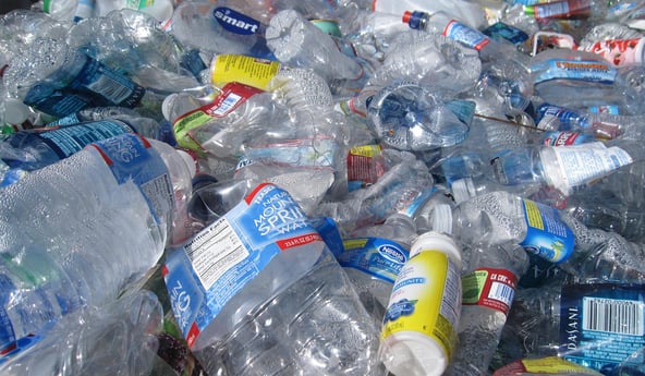
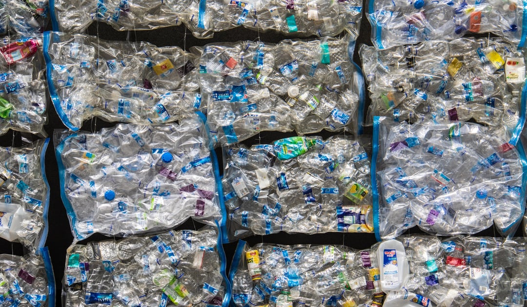
CL imaging in micro- and nanoplastics studies
First time produced in 1907 (1), synthetic plastic is now an integral part of modern life. Plastics owe their success and widespread to their great characteristics. Unfortunately, easy degradation is not one of them. According to the UN environment program "Today, we produce about 300 million tonnes of plastic waste every year. That’s nearly equivalent to the weight of the entire human population" (2).
A large part of this plastic waste disintegrates and forms plastic particles of sizes ranging from nano- to micrometers, called respectively nanoplastics and microplastics. There is a lot of evidence that these particles have detrimental effects on living organisms.
Cathodoluminescence imaging might potentially be an excellent method to further deepen our understanding of the properties of these objects. In comparison with other available techniques, such as optical microscopy, gas chromatography, Raman and FTIR spectroscopy, and SEM-EDX, cathodoluminescence imaging is faster and not too expensive, it gives high-resolution data, can be used to study both nano- and microplastics and can differentiate even between materials of similar elemental composition. It is also compatible with other techniques that provide complementary insights.
We have tested how well CL imaging characterizes various micro and nanoplastic particles. In the study, we used polystyrene beads of 8 µm and 600 nm, as well as polyethylene and polypropylene particles. Using CL imaging, we have acquired spectra characteristics for each of the materials. Moreover, in a sample containing a mix of materials, based on the CL spectra we were able to detect polystyrene and Teflon. These results demonstrate that CL imaging is a promising fast and high-resolution technique to help characterize micro- and nanoplastic particles.
Learn more about this study from our application note.
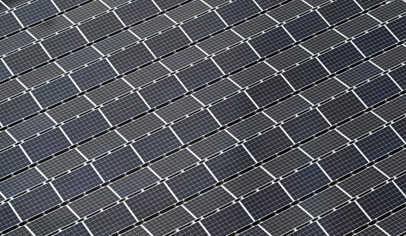
CL imaging in photovoltaics (PV)
The sunlight that hits the Earth in one hour carries more energy than all humans use in a year (3, 4). If we were only able to harvest, convert and store even a small part of this energy efficiently, we could stop producing it from fossil fuels and nuclear fission. The developments in photovoltaics (PV) make it already possible, though mostly on a local scale, to produce affordable and sustainable energy using solar power as an energy source. One of the major challenges in advancing solar cells, however, is improving the efficiency with which they convert sunlight energy into chemical energy.
PV technology relies on complex materials that are combined in a precise spatial arrangement to provide desired optoelectrical properties. CL imaging is a suitable method to characterize these properties of the resulting solar cells with high resolution. As an example, our team performed a study on a thin-film solar cell containing Cu(In, Ga)Se2 absorbers that show record-high energy conversion efficiencies of almost 23%. With point to point detection distance of about 50 nm, one can clearly see how the optoelectrical properties change within and between different layers. For example, it is possible to characterize the local band-gap energy distribution.
Find out how we did the measurements and what other results we obtained in this application note.
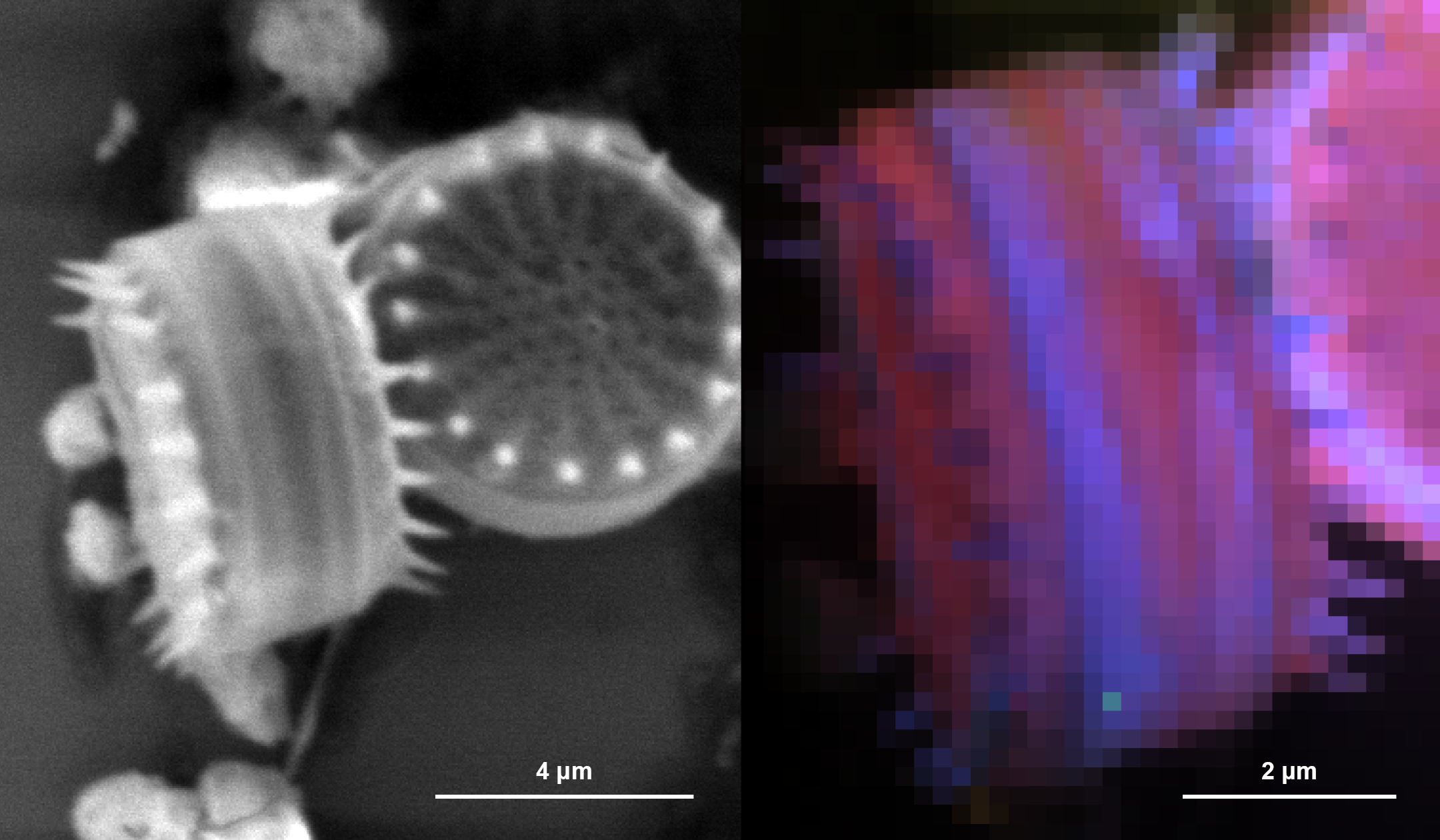
CL imaging in marine microbiology
So far we've shown examples of CL imaging applications in inorganic matter research. However, inspired by the CL studies of geological samples such as silica and carbonate-based materials, we decided to test how well CL imaging will cope with single-cell marine microorganisms. These organisms play a critical role in the Earth's biogeochemical cycles and are tightly interlaced with climate changes (5, 6), therefore marine microbiology is an important subject in environmental sciences. In our study, we applied CL imaging to marine bacteria and unicellular phytoplankton. As a result, based on the CL spectral bands we were able, for example, to distinguish between the silica-containing and carbonate-containing microorganisms and get interesting contrasts at their sub-cellular length scales.
Check our application note on this topic for more details.
These three CL applications in environmental studies are just a few examples of how microscopy-based research supports developments toward a greener and healthier future. Check out the pages for CL imaging in micro- and nanoplastics, PV materials, and marine microorganisms studies and stay up to date with further developments in this important technique.
References
- Our World in Data – Plastic Pollution
- UN Environmental Programme – Beat Plastic Pollution
- Hammarström & Hammes-Schiffer, Artificial Photosynthesis and Solar Fuels Acc. Chem. Res. (2009)
- Scitable by Nature Education – Solar Energy
- Murillo et al., Editorial: Marine Microbiome and Biogeochemical Cycles in Marine Productive Areas Front. Mar. Sci. (2019)
- York, Marine biogeochemical cycles in a changing world Nature (2018)
.gif?width=227&name=CL%20FAQ_Animated%20Banner%20(1).gif)


