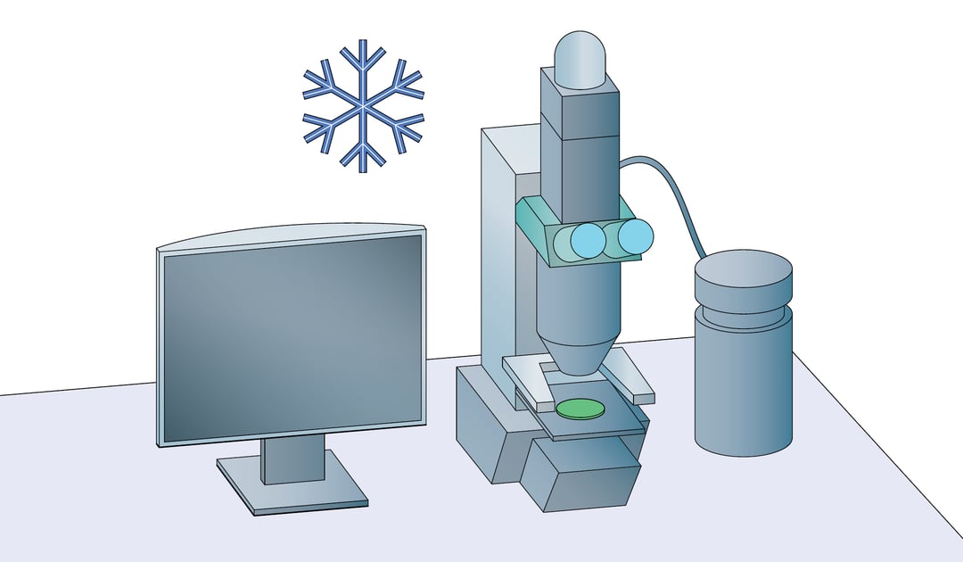
In the cryo-EM workflow, fluorescent light microscopy (FLM) is often used to identify regions of interest (ROIs) for image acquisition. In this context, FLM is performed after vitrification to ensure the highest correlation accuracy between FLM and EM images. This means that fluorescent imaging is also carried out under cryogenic conditions (cryo-FLM). So in what ways is fluorescence imaging different under cryogenic conditions compared to ambient temperatures? What are the technical limitations, and how to overcome them?
Technical limitations of cryo-FLM
Cryo-FLM is rarely used as an independent imaging technique but is mainly used for correlative studies. The main reason for this is that cryo-FLM has a lower effective resolution than FLM at ambient temperatures. This is because cryogenic conditions inhibit the use of immersion objectives. In light microscopy, the high numerical aperture (NA) of immersion objectives allows imaging at resolutions near the diffraction limit at ~200nm. In contrast, the air objectives that are used in cryo-FLM are limited to a lower NA and are therefore only able to reach resolutions of 400 to 500nm.
What to consider when using cryo-FLM in correlative imaging
Cryogenic conditions change many aspects of FLM. Fluorophores behave differently at very low temperatures, leading to some benefits as well as some challenges for cryo-FLM. A major advantage is that under cryogenic conditions, fluorophore bleaching is severely reduced [1]. The process of photobleaching relies on molecular alterations of the fluorophore as well as interactions with
surrounding molecules. Both of these mechanisms are considerably impaired at low temperatures. This means that long and high-intensity exposures can be used during image acquisition.
Another benefit is that most fluorophores have narrower emission and absorption spectra at low temperatures, reducing spectral overlap and allowing better multicolor imaging [2]. A disadvantage is that under cryogenic conditions, fluorophores are far more likely to be excited to a dark state, from which they are not able to emit fluorescence [3]. This results in a significant decrease in the overall signal from the sample. However, these characteristics are not known for all fluorophores and more research is being done to optimize fluorophores for cryogenic imaging.
What is the future of cryo-FLM?
The relatively low effective resolution of cryo-FLM is the largest barrier to its use as a separate technique. Some advances have been made in the design of immersion objectives and immersion fluids that work at very low temperatures [4]. This would bring cryo-FLM to the same level as ambient fluorescence microscopy. Another exciting observation is that the “blinking” of fluorophores still
occurs under cryogenic conditions, allowing cryogenic super-resolution microscopy [5]. These and other studies further increase the significance of cryo-FLM in correlative studies and also pave the way to making cryo-FLM an important independent technique.
Cryo-FLM is an important technique used in the cryo-electron tomography (cryo-ET) workflow. In this workflow an ROI is identified using cryo-FLM, the sample is thinned by a focussed ion beam in a FIB/SEM and finally transferred to a transmission electron microscope (TEM) for imaging. Transferring the delicate sample between the microscopes while keeping it at cryogenic temperatures makes the workflow an exceedingly challenging task.
At Delmic we have developed a solution that integrates cryo-FLM into a cryo-FIB/SEM, thereby reducing the number of handling steps and increasing the yield of the cryo-ET workflow. If you would like to learn more about it, sign up for the updates!
References
[1] C. L. Schwartz, V. I. Sarbash, F. I. Ataullakhanov, J. R. Mcintosh, and D. Nicastro, “Cryo-fluorescence microscopy facilitates correlations between light and cryo-electron microscopy and reduces the rate of photobleaching,” J. Microsc., vol. 227, no. 2, pp. 98–109, 2007, doi:10.1111/j.1365-2818.2007.01794.x.
[2] T. M. H. Creemers, A. J. Lock, V. Subramaniam, T. M. Jovin, and S. Volker, “Three
photoconvertible forms of green fluorescent protein identified by spectral hole-burning,” Nat. Struct. Biol., vol. 6, no. 6, pp. 557–560, 1999, doi: 10.1038/9335.
[3] R. Kaufmann, C. Hagen, and K. Grünewald, “Fluorescence cryo-microscopy: Current challenges and prospects,” Current Opinion in Chemical Biology, vol. 20, no. 1. Elsevier Ltd, pp. 86–91, Jun-2014, doi: 10.1016/j.cbpa.2014.05.007.
[4] R. Faoro et al., “Aberration-corrected cryoimmersion light microscopy,” Proc. Natl. Acad. Sci. U. S. A., vol. 115, no. 6, pp. 1204–1209, Feb. 2018, doi: 10.1073/pnas.1717282115.
[5] M. W. Tuijtel, A. J. Koster, S. Jakobs, F. G. A. Faas, and T. H. Sharp, “Correlative cryo super-resolution light and electron microscopy on mammalian cells using fluorescent proteins,” Sci. Rep., vol. 9, no. 1, pp. 1–11, Dec. 2019, doi:10.1038/s41598-018-37728-8.
This work is supported by the European SME2 grant № 879673 - Cryo-SECOM Workflow
.png)


