.svg "DELMIC")
.png)


In this blog post, we highlight a study in which researchers applied in situ cryo-electron tomography to uncover several previously unknown characteristics of molecular organization and interactions between main proteins in skeletal muscles.
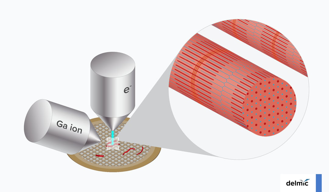
The basic contractile unit in a skeletal muscle tissue is called a sarcomere. The two main proteins contained in a sarcomere, actin and myosin, form long parallel filaments that slide past each other causing the muscle to contract or relax. Even though sarcomeres and some of their components were characterized already in the 1800s, it wasn't until the 1950s that this sliding filament model was used to explain how sarcomeres generate force and bear load, thus supporting the musculoskeletal functions (3). Further understanding of how sarcomers work was possible thanks to the use of, for example, X-ray diffraction, light microscopy and electron microscopy (EM). But these traditional techniques have often suffered from artifacts caused by sample preparation and the resulting lower resolution. To circumvent these drawbacks, Wang et al. from the Raunser group at Max Planck Institute of Molecular Physiology in Dortmund, Germany, applied in their studies in situ cryo-electron tomography (cryo-ET) (4). Thanks to this approach, they were able to provide several new insights about the 3D molecular organization and interactions between the major sarcomeric proteins in a mouse psoas muscle. One of the most important findings in this work is that the myosin "double-head" can interact not only with the same actin filament, but sometimes it actually splits the heads between two actin filaments. Even though previous studies speculated the existence of such a "split-head" arrangement, this was the first direct proof of it. The visualized underlying conformation of myosin revealed moreover the ability of this molecule to accommodate force production, providing a great example of how structural information with a molecular detail can uncover protein's mechanism.
To obtain these insights, the researchers performed segmentation of acquired cryo-ET tomograms, followed by sub-volume averaging of various regions of interest. As a result, the cross-bridges formed between myosin heads and actin were reconstructed at 10.2 Å resolution, which enabled the fitting of the density maps with adequate cryo-EM and crystal structures.
By applying this experimental approach, Wang and colleagues gained new insights also in two other sarcomeric proteins, tropomyosin and α-actinin. The former is a double-stranded α-helical coiled protein which winds around an actin filament and plays a crucial role in regulating its function. α-actinin, on the other hand, cross-links actin filaments in the end-regions of a sarcomere and the interaction of α-actinin with other proteins is particularly interesting with respect to diseases such as actininopathies, zaspopathies, nemaline myopathies and myofibrillar myopathies.
In 2005 there were 20 known skeletal muscle diseases linked to mutations in sarcomeric proteins (2), a number likely much higher now, after 16 years of intense medical research and advancements in omics and bioinformatics. In the foreseeable future, further developments in cryo-ET will undoubtedly enable researchers to shed light on the molecular basis of many of these conditions and pave a way for future medical treatments. It is exciting to observe the progress of this technology, but even more exciting for us at Delmic is to actively contribute to this momentum by developing solutions that let the researchers get there faster.
References
[1] https://www.who.int/news-room/fact-sheets/detail/musculoskeletal-conditions
[2] Laing and Nowak, When contractile proteins go bad: the sarcomere and skeletal muscle disease BioEssays (2005) - link
[3] Squire, Muscle contraction: Sliding filament history, sarcomere dynamics and the two Huxleys Global Cardiology Science and Practice (2016) - link
[4] Wang et al., The molecular basis for sarcomere organization in vertebrate skeletal muscle Cell (2021) - link
This work is supported by the European SME2 grant № 879673 - Cryo-SECOM Workflow.