Cryogenic electron tomography (cryo-ET) is an emerging powerful technique that can resolve biological structures such as intracellular organelles and protein complexes in their near-native state at a high resolution. One of the most challenging steps in the cryo-ET workflow is in lamella preparation. Biological samples need to be 150-300 nm thick to be electron transparent. Cellular and multicellular samples can be focused ion beam (FIB) milled to generate lamellae of the required thickness range. To find the region of interest (ROI) cryo-correlative light and electron microscopy (cryo-CLEM) has proven to be useful. The use of a separate cryo-fluorescent light microscope (cryo-FLM) does however pose the risk of damage, contamination and devitrification of the precious samples.
An improved cryo-ET workflow
To overcome these challenges, Delmic developed METEOR: a FLM that can be integrated into a cryo-FIB/SEM system. Having a FLM inside the cryo-FIB/SEM greatly reduces the amount of handling steps required, resulting in less contamination, less damage and a more efficient cryo-ET workflow. Additionally, switching between FLM and FIB/SEM imaging without moving the sample significantly speeds up the identification of ROIs. Furthermore, it is easy to reinspect the sample with FLM during and after the milling process, thereby improving the quality of the samples that ultimately end up in the cryo-TEM.
Guided lamella milling of yeast and HeLa cells
We have shown that METEOR can streamline the cryo-ET workflow for targeted lamella milling of HeLa and yeast cells. HeLa cells and yeast cells are important model systems used to study cellular processes that have contributed to many medical breakthroughs.
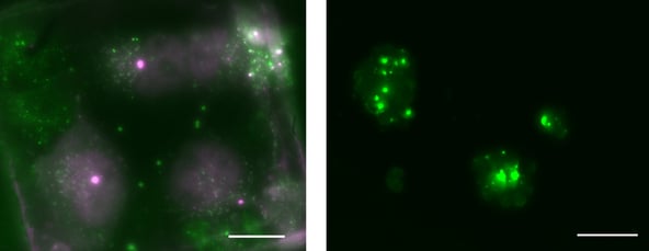
We used a HeLa cell line in which two proteins were tagged: a protein localizing to mitochondria was tagged with eGFP and the second protein of interest was tagged with mCherry. The region where the two proteins colocalized was our ROI. In the second test case, we used yeast cells that overexpressed Ede1 tagged with eGFP. The locations where Ede1 clustered was our ROI for this sample.

Left: dual colour HeLa cells Green: protein localizing on the mitochondria Pink: second protein of interest (scale bar: 20 μm). Right: METEOR images of yeast cells overexpressing Ede1-eGFP. Images courtesy of A. Bieber, C. Capitanio and O. Schioetz (MPI Biochemistry, Germany)
We took METEOR images before milling and correlated them with FIB images to guide lamella milling. During milling, METEOR images were taken to confirm that the current milling strategy was correct. After milling we took a final METEOR image to confirm the ROI was still present in the lamella.
Future possibilities of integrated cryo-FLM
We have shown how METEOR can streamline the milling of HeLa and yeast cells. Since METEOR is a very flexible system it can be applied to a wide range of research areas and workflows. METEOR could also be used to enhance research areas such as virology, membrane trafficking and neurobiology. In addition, METEOR could also be used in automated milling workflows or cryo-FIB lift-out.
Do you want to save this blog in PDF? Download the flyer here. To learn more about METEOR and its applications, we recommend you to read its application note below!
This work is supported by the European SME2 grant № 879673 - Cryo-SECOM Workflow.
.png)


.png?width=1080&name=2021_HeLA%20Yeast%20Blogpost_Website%20(1).png)


