materials science
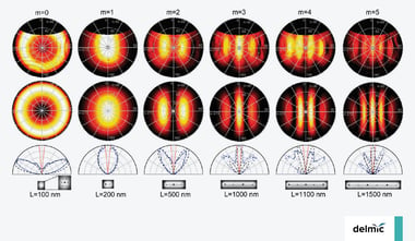
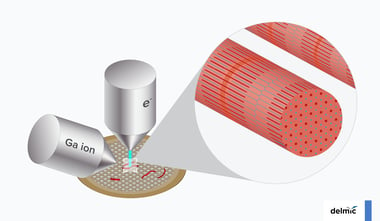
Angle-resolved cathodoluminescence imaging for nanophotonics
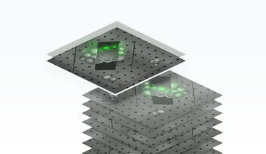
The advancement of nanotechnology opened up exciting new possibilities to create nanophotonic structures with nanoscale dimensions. A fundamental understanding ...
.svg "DELMIC")
.png)
Acquire more powerful insights to progress your research
Resolve optical properties at the nanoscale


All topics
The advancement of nanotechnology opened up exciting new possibilities to create nanophotonic structures with nanoscale dimensions. A fundamental understanding ...

Traditionally, scanning electron microscopy (SEM) data management has involved data transfer from a microscope to a hard disc and then loading the data to a ...
Cryogenic fluorescence microscopy (cryo-FLM) combined with scanning electron microscopy (SEM) makes the process of lamella milling in the cryo-electron ...
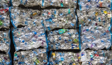
Understanding how different materials work and knowing their properties has been a keystone in human progress. In our efforts to build an environmentally ...
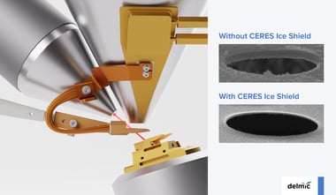
Sample milling is a crucial step in the Cryo-ET workflow. Unfortunately, this step poses yet another occasion for ice contamination to derail efforts to obtain ...

With more than 90 years of development, electron microscopy is now a mature, well-established technique applied in many life science fields, such as pathology, ...
There are more than 150 known musculoskeletal conditions (1), many of which affect specifically skeletal muscles and can lead to lifelong disabilities or even ...
Cryo-EM research helps us understand how the world works on a microscale. Often these insights have a great impact on our lives, for example, the understanding ...
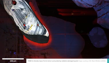
Geochronology plays a central role in geosciences and all historical aspects of the Earth sciences. Over the years, it has become an essential scientific field ...
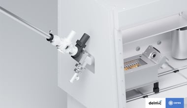
Sample transfer is definitely not the most spectacular part of the cryo-ET workflow. And yet, it is one of the most problematic steps, disturbing many ...
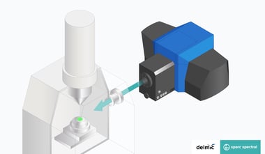
Cathodoluminescence (CL) is the process of light emission from a material as a result of excitation by electrons. Imaging CL can therefore enable us to study ...
Cryo-electron tomography (cryo-ET) is an extremely powerful technique that allows studies of the cellular landscape at high resolution in a near-native state.