
life sciences
Public Image Archives: The Power of Data Sharing for 3D EM
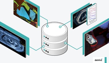
Although most scientific articles impose copyright regulations to prevent sharing and using their 3D electron microscopy (EM) data, public image archives ...
.svg "DELMIC")
.png)
Acquire more powerful insights to progress your research
Resolve optical properties at the nanoscale


All topics
Although most scientific articles impose copyright regulations to prevent sharing and using their 3D electron microscopy (EM) data, public image archives ...
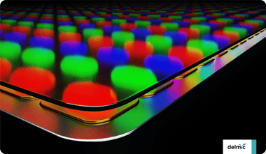
Prominent consumer electronics and LED companies, including Apple, are now developing microLED technology. Samsung has already launched microLED TVs. However, ...
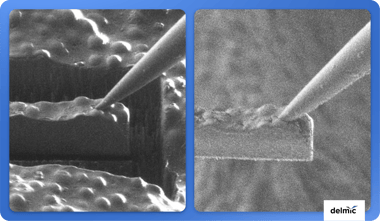
As the boundaries of cryo-electron tomography are continuously being pushed, there are major advancements in the development of cryo-FIB lift-out techniques, ...
The analysis of rock samples and minerals in structural geology is now often being done using luminescence techniques. In this blog post, we will highlight the ...

Artificial intelligence (AI) is increasingly used in life sciences, making communication between these fields crucial. Here, we will highlight community ...
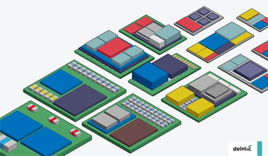
The roadmap for the semiconductor industry has shifted from being solely based on Moore's law to incorporating various focus areas, such as 'More than Moore'. ...
Array tomography, which is increasingly used for producing high-resolution 3D images of tissue samples, includes cutting the sample into ultrathin sections. ...
Many of Delmic’s products come with ODEMIS, our open-source microscopy software for image acquisition and analysis. Here, we will highlight the advantages of ...
Electron microscopy (EM) is a powerful tool for life sciences, with the emergence of volume electron microscopy (volume-EM) enabling 3D imaging of samples with ...
Advancements in technology increased the popularity of cryo-electron microscopy (cryo-EM) techniques such as cryo-electron tomography (cryo-ET) and ...
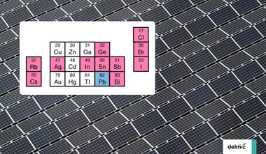
Perovskite solar cells could spur the growth of solar power, but they currently contain lead, which is toxic. Recently, scientists have started to explore ...
We are moving towards a future of open source scientific publications. Apart from publications, software can also be ‘closed’ or ‘open’ source. What does this ...