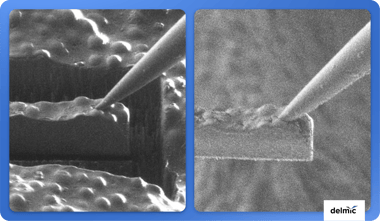
DELMIC
Delmic_Black+Blue(preferred)@2x(1)
Twitter-logo Linkedin-logo Youtube-logo
  • Life Sciences arrow image

    Delmic for life sciences

    Acquire more powerful insights to progress your research

    Explore solutions

    Our solutions

    • product-icon_METEOR METEOR
    • product-icon_FAST-EM FAST-EM
    • product-icon_CERES-IS CERES Ice Shield
    • product-icon_CERES-CS-VL CERES Sample Preparation
    • product-icon_SECOM SECOM

    Workflows

    • Cryo Electron Tomography
    • High-throughput EM
    • Correlative light EM

    Insights

    • Publications
    • Blog
    • Webinars
    • White papers
    • Application notes
    • Technical notes
  • Materials Analysis arrow image

    Delmic for materials analysis

    Resolve optical properties at the nanoscale

    Explore solutions

    Our solutions

    • product-icon_SPARC-Spectral SPARC Spectral
    • product-icon_SPARC Compact SPARC Compact
    • product-icon_JOLT JOLT 2.0
    • product-icon_LAB-Cube LAB Cube

    Techniques

    • Cathodoluminescence

    Applications

    • Geology
    • Semiconductors
    • Nanophotonics
    • Quantum

    Insights

    • Publications
    • Blog
    • Webinars
    • White papers
    • Application notes
    • Technical notes
  • Why Delmic?
  • Insights arrow image

    Insights

    • Blog
    • Customer stories
    • Resource library
    • Feed

    Technique Deep Dives

    • Cathodoluminescence
    • Cryo-electron tomography
    • High-throughput EM
    • Correlative light and electron microscopy
  • Company arrow image

    What do we do?

    We aspire to achieve seamless microscopy as the standard

    Learn more

    Company

    • About us
    • News
    • Career
    • Distributors
    • Contact
Search
Get in touch
Search
hamburger close

Blog

  • life sciences
  • cathodoluminescence
  • cryo electron microscopy
  • cryo-electron tomography
  • SPARC
  • materials science
  • fast SEM imaging
  • FAST-EM
  • geology
  • nanophotonics
  • correlative light and electron microscopy
  • semiconductors
  • SECOM
  • METEOR
  • cryo-focused ion beam
  • time-resolved cathodoluminescence
  • photovoltaics
  • software
  • volume electron microscopy
  • Array tomography
  • State of electron microscopy
  • plasmonics
  • History of microscopy
  • LAB Cube
  • Scanning electron microscope (SEM)
  • artificial intelligence
  • drug development
  • event
  • microplastics
  • power electronics
  • webinar
  • 冷冻电子断层扫描
  • All topics

life sciences
Community Platforms to Advance Life Science Research Using AI

Artificial intelligence (AI) is increasingly used in life sciences, making communication between these fields crucial. Here, we will highlight community ...

life sciences
Array tomography workflow: tips and tricks on section handling from researchers in the field

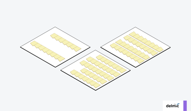
Array tomography, which is increasingly used for producing high-resolution 3D images of tissue samples, includes cutting the sample into ultrathin sections. ...

life sciences
What is the future of electron microscopy for life sciences?

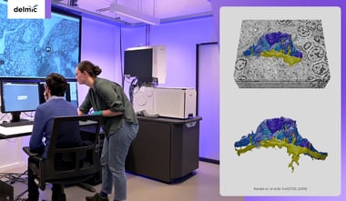
Electron microscopy (EM) is a powerful tool for life sciences, with the emergence of volume electron microscopy (volume-EM) enabling 3D imaging of samples with ...

life sciences
What is the difference between cryo-EM and cryo-ET?

Advancements in technology increased the popularity of cryo-electron microscopy (cryo-EM) techniques such as cryo-electron tomography (cryo-ET) and ...

life sciences
How electron microscopy shapes the way we think, according to 6 prominent scientists

life sciences
A molecular view on Parkinson’s disease using cryo-EM

Advances in cryo electron microscopy made it possible to shed light on the molecular pathology of Parkinson's disease. Here, we discuss recent exciting ...

life sciences
Rubisco assembly: the promise of cryo-ET for synthetic biologists

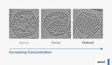
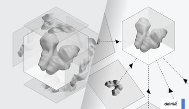
In this blog post, we discuss the ultrastructure and assembly mechanisms of Rubisco, revealed by cryo-ET. Together with advances in synthetic biology, these ...

life sciences
Nanoscale Resolution Connectomes: Pushing the Limits of Brain Imaging

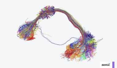
With advancements in electron microscopy, researchers now aim to build connectomes of increasingly larger brain volumes. In this blog, we discuss exciting ...

life sciences
Nanoscale 3D organoid imaging: a powerful tool for drug development

Organoids enhance drug screening methods by mimicking in vivo organs. In this blog, we will discuss the exciting possibilities of 3D electron microscopy ...

life sciences
Revealing the ultrastructure of autophagic membranes during different stages of biogenesis with correlative cryo-ET

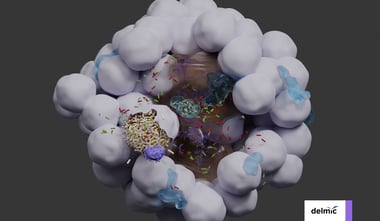
By visualizing membrane ultrastructure directly in the cells, researchers can better understand their diversity, dynamics, and role. In this blog post, we ...

life sciences
Volume Electron Microscopy: Towards Automatic Organelle Segmentation

Manual segmentation of organelles hampers the use of volume electron microscopy in biological research. In this blog, we will discuss a novel approach to ...

life sciences
Advancing pathology with 3D high-throughput nanoscale imaging

Using electron microscopy (EM), pathologists can visualize ultrastructural details in biological samples. In this blog, we discuss how recent developments in ...

  • 1
  • 2
  • 3
  • →
Delmic_Black+Blue(preferred)@2x

Life science Frame 264(3)

Solutions
  • METEOR
  • FAST-EM
  • CERES Ice Shield
  • CERES Sample Preparation
  • SECOM
Workflows
  • Cryo Electron Tomography
  • High-throughput EM
  • Correlative light EM

Materials analysis Frame 264(3)

Solutions
  • SPARC Spectral
  • SPARC Compact
  • JOLT 2.0
  • LAB Cube
Techniques
  • Cathodoluminescence
Applications
  • Geology
  • Semiconductors
  • Nanophotonics
  • Quantum

Insights Frame 264(3)

  • Blog
  • Customer stories
  • Resource library
Deep dive
  • Cathodoluminescence
  • Cryo-EM
  • High-throughput EM
  • CLEM

Company Frame 264(3)

  • About Delmic
  • News
  • Distributors
  • Careers
  • Contact