life sciences
Preventing ice contamination in the cryo-ET workflow
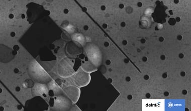
One of the biggest hurdles in the current cryo-ET workflow is avoiding ice contamination of the vitreous sample, which is difficult to avoid due to the many ...
.svg "DELMIC")
.png)
Acquire more powerful insights to progress your research
Resolve optical properties at the nanoscale


All topics
One of the biggest hurdles in the current cryo-ET workflow is avoiding ice contamination of the vitreous sample, which is difficult to avoid due to the many ...
Cryo-electron tomography (cryo-ET) is an extremely powerful technique that allows studies of the cellular landscape at high resolution in a near-native state. ...
Parkinson's disease (PD) is a neurodegenerative disease that mainly affects the motor system and its functioning. About one tenth of patients with PD has ...
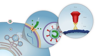
This year was dominated by the outbreak of SARS-CoV-2. Around the world, many researchers are looking into finding a solution for the highly pathogenic ...
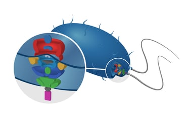
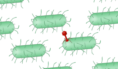
Flagella are filamentous protein complexes found on many bacteria and some eukaryotic cells. Imaging techniques such as cryo-electron tomography (cryo-ET), are ...
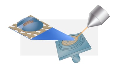
The development of high throughput electron microscopy techniques in the last decade has made increasingly large imaging projects possible. Now that systems ...
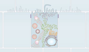
Eukaryotic cells contain a complex network of membrane-bound organelles where specialized processes of the cell take place.
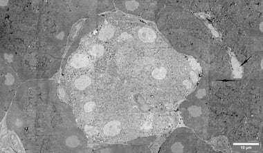
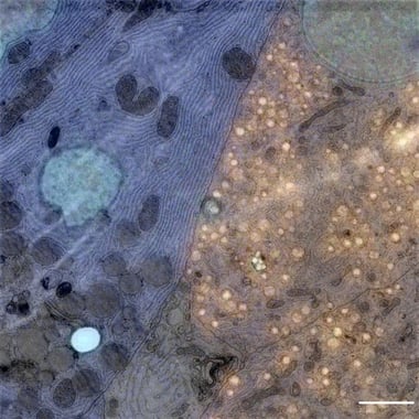
Comparing the morphology of healthy and diseased cells or tissues or examining the effect of drug treatments is extremely important for understanding of the ...
Viruses are microscopic infectious agents that are not able to reproduce outside of their hosts. We associate viruses with a variety of diseases, ranging from ...
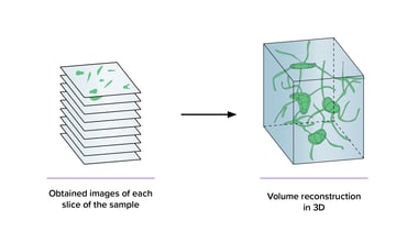
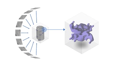
As we all know, biological creatures are highly complex and three-dimensional. Therefore, it is crucial to capture 3D images of their cell structures to find ...
Over the past decades, researchers have been dedicated to studying the beta cells in the islets of Langerhans in order to better understand Diabetes Type 1, ...
Understanding the relationship between structure and function in biology requires continuous developments in the field of microscopy.