.svg "DELMIC")
.png)


life sciences
How 3D Cellular Atlases and Volume EM Are Transforming Biological Discovery
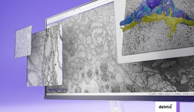
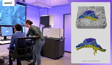
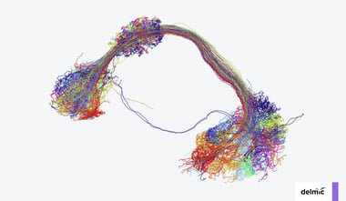
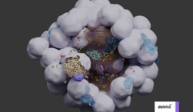
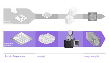
Large-scale volume electron microscopy (vEM) is transforming biological research by making it possible to image cells and tissues in 3D at nanoscale resolution. This combination of scale and detail...