.svg "DELMIC")
-
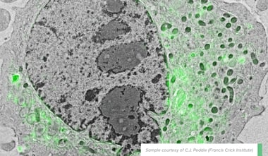
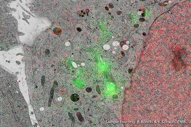
Life Sciences
.png)
Delmic for life sciences
Acquire more powerful insights to progress your research
Our solutions
-
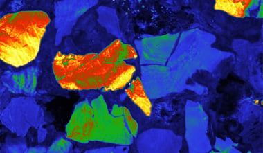
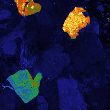
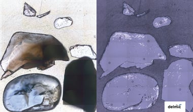
Materials Analysis
Delmic for materials analysis
Resolve optical properties at the nanoscale
Our solutions
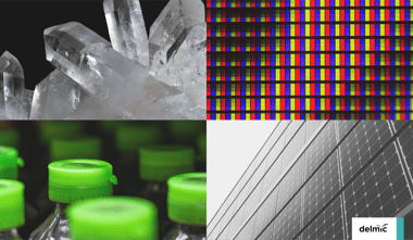
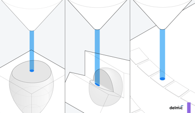
Techniques
Applications
- Why Delmic?
-
Insights
-
Company


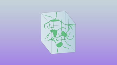
.png?width=380&name=2021_HeLA%20Yeast%20Blogpost_Website%20(1).png)